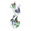
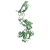
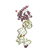
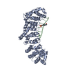
[English] 日本語
Yorodumi
- PDB-2p26: Structure of the PHE2 and PHE3 fragments of the integrin beta2 subunit -

+
Open data


ID or keywords:

Loading...

-
Basic information

Entry
Database: PDB / ID: 2p26
TitleStructure of the PHE2 and PHE3 fragments of the integrin beta2 subunit
ComponentsIntegrin beta-2
KeywordsCELL ADHESION / Integrin Beta2 Subunit / hybrid domain / PSI domain / I-EGF domains
Function / homology
Function and homology information


integrin alphaX-beta2 complex / integrin alphaM-beta2 complex / positive regulation of prostaglandin-E synthase activity / positive regulation of neutrophil degranulation / integrin alphaL-beta2 complex / ICAM-3 receptor activity / complement component C3b binding / neutrophil migration / Toll Like Receptor 4 (TLR4) Cascade / negative regulation of dopamine metabolic process ...integrin alphaX-beta2 complex / integrin alphaM-beta2 complex / positive regulation of prostaglandin-E synthase activity / positive regulation of neutrophil degranulation / integrin alphaL-beta2 complex / ICAM-3 receptor activity / complement component C3b binding / neutrophil migration / Toll Like Receptor 4 (TLR4) Cascade / negative regulation of dopamine metabolic process / cell-cell adhesion via plasma-membrane adhesion molecules / heterotypic cell-cell adhesion / integrin complex / positive regulation of leukocyte adhesion to vascular endothelial cell / leukocyte cell-cell adhesion / phagocytosis, engulfment / receptor clustering / amyloid-beta clearance / endodermal cell differentiation / plasma membrane raft / tertiary granule membrane / ficolin-1-rich granule membrane / cellular response to low-density lipoprotein particle stimulus / positive regulation of protein targeting to membrane / Integrin cell surface interactions / specific granule membrane / regulation of peptidyl-tyrosine phosphorylation / heat shock protein binding / receptor-mediated endocytosis / cell adhesion molecule binding / cell-matrix adhesion / neutrophil chemotaxis / positive regulation of superoxide anion generation / integrin-mediated signaling pathway / Cell surface interactions at the vascular wall / microglial cell activation / receptor internalization / cell-cell adhesion / extracellular vesicle / Immunoregulatory interactions between a Lymphoid and a non-Lymphoid cell / integrin binding / cell-cell signaling / amyloid-beta binding / regulation of cell shape / Interleukin-4 and Interleukin-13 signaling / receptor complex / cell adhesion / inflammatory response / external side of plasma membrane / focal adhesion / apoptotic process / Neutrophil degranulation / protein kinase binding / cell surface / extracellular exosome / membrane / metal ion binding / plasma membrane
Similarity search - Function
Integrin beta-2 subunit / ligand-binding face of the semaphorins, domain 2 / ntegrin, alpha v. Chain A, domain 3 / ligand-binding face of the semaphorins, domain 2 / Integrin beta subunit, cytoplasmic domain / Integrin beta cytoplasmic domain / Integrin_b_cyt / Integrin beta tail domain / Integrin beta subunit, tail / Integrin beta tail domain superfamily ...Integrin beta-2 subunit / ligand-binding face of the semaphorins, domain 2 / ntegrin, alpha v. Chain A, domain 3 / ligand-binding face of the semaphorins, domain 2 / Integrin beta subunit, cytoplasmic domain / Integrin beta cytoplasmic domain / Integrin_b_cyt / Integrin beta tail domain / Integrin beta subunit, tail / Integrin beta tail domain superfamily / Integrin_B_tail / Integrin beta subunit, VWA domain / Integrin beta subunit / Integrin beta N-terminal / Integrin beta chain VWA domain / Integrin plexin domain / Integrins beta chain cysteine-rich domain signature. / Integrin beta subunits (N-terminal portion of extracellular region) / Integrin domain superfamily / PSI domain / domain found in Plexins, Semaphorins and Integrins / Laminin / Laminin / von Willebrand factor A-like domain superfamily / EGF-like domain signature 2. / EGF-like domain signature 1. / Ribbon / Immunoglobulin-like / Sandwich / 2-Layer Sandwich / Mainly Beta / Alpha Beta
Similarity search - Domain/homology
Biological speciesHomo sapiens (human)
MethodX-RAY DIFFRACTION / MOLECULAR REPLACEMENT / Resolution: 1.75 Å
AuthorsShi, M. / Foo, S.Y. / Tan, S.M. / Mitchell, E.P. / Law, S.K.A. / Lescar, J.
CitationJournal: J.Biol.Chem. / Year: 2007
Title: A structural hypothesis for the transition between bent and extended conformations of the leukocyte beta2 integrins
Authors: Shi, M. / Foo, S.Y. / Tan, S.M. / Mitchell, E.P. / Law, S.K.A. / Lescar, J.
History
DepositionMar 6, 2007Deposition site: RCSB / Processing site: PDBJ
Revision 1.0Aug 14, 2007Provider: repository / Type: Initial release
Revision 1.1Jul 13, 2011Group: Advisory / Version format compliance
Revision 1.2Aug 16, 2017Group: Refinement description / Source and taxonomy / Category: entity_src_gen / software
Revision 1.3Jul 29, 2020Group: Data collection / Database references ...Data collection / Database references / Derived calculations / Structure summary
Category: chem_comp / entity ...chem_comp / entity / pdbx_chem_comp_identifier / pdbx_entity_nonpoly / struct_conn / struct_ref_seq_dif / struct_site / struct_site_gen
Item: _chem_comp.name / _chem_comp.type ..._chem_comp.name / _chem_comp.type / _entity.pdbx_description / _pdbx_entity_nonpoly.name / _struct_conn.pdbx_leaving_atom_flag / _struct_conn.pdbx_role / _struct_conn.ptnr1_auth_comp_id / _struct_conn.ptnr1_auth_seq_id / _struct_conn.ptnr1_label_asym_id / _struct_conn.ptnr1_label_atom_id / _struct_conn.ptnr1_label_comp_id / _struct_conn.ptnr1_label_seq_id / _struct_conn.ptnr2_auth_comp_id / _struct_conn.ptnr2_auth_seq_id / _struct_conn.ptnr2_label_asym_id / _struct_conn.ptnr2_label_atom_id / _struct_conn.ptnr2_label_comp_id / _struct_conn.ptnr2_label_seq_id / _struct_ref_seq_dif.details
Description: Carbohydrate remediation / Provider: repository / Type: Remediation
Revision 1.4Oct 25, 2023Group: Data collection / Database references ...Data collection / Database references / Refinement description / Structure summary
Category: chem_comp / chem_comp_atom ...chem_comp / chem_comp_atom / chem_comp_bond / database_2 / pdbx_initial_refinement_model
Item: _chem_comp.pdbx_synonyms / _database_2.pdbx_DOI / _database_2.pdbx_database_accession

-
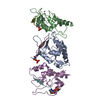
Structure visualization

Structure viewerMolecule:
MolmilJmol/JSmol

Downloads & links

-
Assembly

Deposited unit
A: Integrin beta-2
hetero molecules


Theoretical massNumber of molelcules
Total (without water)31,7244
Polymers31,0601
Non-polymers6643
Water4,828268
1


  • Idetical with deposited unit
  • defined by author&software
TypeNameSymmetry operationNumber
identity operation1_555x,y,z1
MethodPISA
Unit cell
Length a, b, c (Å)58.489, 30.853, 65.157
Angle α, β, γ (deg.)90.00, 94.29, 90.00
Int Tables number4
Space group name H-MP1211

-
Components

#1: Protein Integrin beta-2 / Cell surface adhesion glycoproteins LFA- 1/CR3/p150 / 95 subunit beta / Complement receptor C3 ...Cell surface adhesion glycoproteins LFA- 1/CR3/p150 / 95 subunit beta / Complement receptor C3 subunit beta / CD18 antigen


Mass: 31060.072 Da / Num. of mol.: 1 / Fragment: PHE2 and PHE3
Source method: isolated from a genetically manipulated source
Source: (gene. exp.) Homo sapiens (human) / Plasmid: pIRES2-EGFP / Cell line (production host): HEK293_Gnti / Production host: Homo sapiens (human) / References: UniProt: P05107
#2: Sugar ChemComp-NAG / 2-acetamido-2-deoxy-beta-D-glucopyranose / N-acetyl-beta-D-glucosamine / 2-acetamido-2-deoxy-beta-D-glucose / 2-acetamido-2-deoxy-D-glucose / 2-acetamido-2-deoxy-glucose / N-ACETYL-D-GLUCOSAMINE / N-Acetylglucosamine


Type: D-saccharide, beta linking / Mass: 221.208 Da / Num. of mol.: 3
Source method: isolated from a genetically manipulated source
Formula: C8H15NO6
IdentifierTypeProgram
DGlcpNAcbCONDENSED IUPAC CARBOHYDRATE SYMBOLGMML 1.0
N-acetyl-b-D-glucopyranosamineCOMMON NAMEGMML 1.0
b-D-GlcpNAcIUPAC CARBOHYDRATE SYMBOLPDB-CARE 1.0
GlcNAcSNFG CARBOHYDRATE SYMBOLGMML 1.0
#3: Water ChemComp-HOH / water / Water


Mass: 18.015 Da / Num. of mol.: 268 / Source method: isolated from a natural source / Formula: H2O

-
Experimental details

-
Experiment

ExperimentMethod: X-RAY DIFFRACTION / Number of used crystals: 1

-
Sample preparation

CrystalDensity Matthews: 1.89 Å3/Da / Density % sol: 34.87 %
Crystal growTemperature: 291 K / Method: vapor diffusion, hanging drop / pH: 4.6
Details: 0.2M Magnesium acetate, 0.1M sodium acetate, pH4.6, 18% PEG3350, VAPOR DIFFUSION, HANGING DROP, temperature 291K

-
Data collection

Diffraction sourceSource: ROTATING ANODE / Type: RIGAKU RU300 / Wavelength: 1.5418 Å
DetectorDate: Aug 26, 2006
RadiationProtocol: SINGLE WAVELENGTH / Monochromatic (M) / Laue (L): M / Scattering type: x-ray
Radiation wavelengthWavelength: 1.5418 Å / Relative weight: 1
ReflectionResolution: 1.75→27.87 Å / Num. obs: 21822

-
Phasing

Phasing MR
Highest resolutionLowest resolution
Rotation2.5 Å27.87 Å
Translation2.5 Å27.87 Å

-
Processing

Software
NameVersionClassificationNB
PHASERphasing
REFMAC5.2.0019refinement
PDB_EXTRACT2data extraction
CrystalCleardata collection
MOSFLMdata reduction
SCALAdata scaling
RefinementMethod to determine structure: MOLECULAR REPLACEMENT
Starting model: PDB ENTRY 1YUK
Resolution: 1.75→27.87 Å / Cor.coef. Fo:Fc: 0.951 / Cor.coef. Fo:Fc free: 0.916 / SU B: 6.87 / SU ML: 0.114 / TLS residual ADP flag: LIKELY RESIDUAL / Cross valid method: THROUGHOUT / σ(F): 0 / ESU R: 0.179 / ESU R Free: 0.161 / Stereochemistry target values: MAXIMUM LIKELIHOOD / Details: HYDROGENS HAVE BEEN ADDED IN THE RIDING POSITIONS
RfactorNum. reflection% reflectionSelection details
Rfree0.253 1117 5.1 %RANDOM
Rwork0.204 ---
obs0.207 21815 91.34 %-
Solvent computationIon probe radii: 0.8 Å / Shrinkage radii: 0.8 Å / VDW probe radii: 1.4 Å / Solvent model: MASK
Displacement parametersBiso mean: 20.372 Å2
Baniso -1Baniso -2Baniso -3
1--0.21 Å20 Å20.2 Å2
2---0.02 Å20 Å2
3---0.26 Å2
Refinement stepCycle: LAST / Resolution: 1.75→27.87 Å
ProteinNucleic acidLigandSolventTotal
Num. atoms2156 0 42 268 2466
Refine LS restraints
Refine-IDTypeDev idealDev ideal targetNumber
X-RAY DIFFRACTIONr_bond_refined_d0.0070.0212281
X-RAY DIFFRACTIONr_angle_refined_deg1.1691.9673095
X-RAY DIFFRACTIONr_dihedral_angle_1_deg5.8175285
X-RAY DIFFRACTIONr_dihedral_angle_2_deg35.74424.22109
X-RAY DIFFRACTIONr_dihedral_angle_3_deg15.03315384
X-RAY DIFFRACTIONr_dihedral_angle_4_deg12.7831518
X-RAY DIFFRACTIONr_chiral_restr0.080.2338
X-RAY DIFFRACTIONr_gen_planes_refined0.0030.021740
X-RAY DIFFRACTIONr_nbd_refined0.1890.2957
X-RAY DIFFRACTIONr_nbtor_refined0.3010.21516
X-RAY DIFFRACTIONr_xyhbond_nbd_refined0.1110.2207
X-RAY DIFFRACTIONr_symmetry_vdw_refined0.1850.2157
X-RAY DIFFRACTIONr_symmetry_hbond_refined0.1330.241
X-RAY DIFFRACTIONr_mcbond_it0.3781.51448
X-RAY DIFFRACTIONr_mcangle_it0.64922275
X-RAY DIFFRACTIONr_scbond_it1.1013907
X-RAY DIFFRACTIONr_scangle_it1.7944.5820
LS refinement shellResolution: 1.75→1.795 Å / Total num. of bins used: 20
RfactorNum. reflection% reflection
Rfree0.592 36 -
Rwork0.404 817 -
obs-853 48.41 %
Refinement TLS params.

Method: refined / Refine-ID: X-RAY DIFFRACTION

IDL112)L122)L132)L222)L232)L332)S11 (Å °)S12 (Å °)S13 (Å °)S21 (Å °)S22 (Å °)S23 (Å °)S31 (Å °)S32 (Å °)S33 (Å °)T112)T122)T132)T222)T232)T332)Origin x (Å)Origin y (Å)Origin z (Å)
12.06580.1150.72792.2788-0.29211.72430.0247-0.04-0.05970.0526-0.00080.0902-0.0941-0.101-0.0240.06220.0117-0.00350.00560.00140.0181-18.898-7.859-32.05
27.8094-1.52932.8792.0915-2.51589.00430.591-0.0352-1.81470.0828-0.2474-0.19950.97721.0782-0.34360.30170.0437-0.07590.33940.10470.464312.032-18.827-11.832
33.9144-0.45090.94981.3659-0.04211.34440.0428-0.4148-0.14060.20240.05560.04130.0195-0.0226-0.09830.0689-0.0193-0.00960.0287-0.006-0.03977.676-7.756-11.073
43.2338-0.66921.30062.6518-1.55018.0144-0.0421-0.63350.25840.29090.23430.096-0.0785-0.2294-0.19210.0431-0.0103-0.00010.0937-0.0809-0.03375.149-2.346-5.144
53.6964-0.13521.52440.3879-0.01321.25950.03940.0895-0.06570.0496-0.03210.0536-0.00290.1464-0.00730.0586-0.0193-0.00660.0098-0.0060.00895.966-6.934-20.77
61.9944-1.891-0.05822.23120.57893.87420.09160.0142-0.0987-0.1132-0.08950.0570.2312-0.0664-0.00210.0737-0.0058-0.0172-0.0050.015-0.0001-16.342-20.196-45.973
76.5756-2.84820.88714.9567-0.55135.18110.18840.35080.412-0.1237-0.2035-0.1579-0.1364-0.04580.01510.05360.0378-0.00750.060.0282-0.0193-20.867-12.589-52.915
83.3571.21232.94536.2386-0.83593.20610.37350.38320.4884-1.18350.1059-0.4308-0.60540.6698-0.47930.26390.0120.09950.20030.06870.0157-29.184-0.722-57.038
93.1521-0.6472-2.98931.80161.17724.31390.13890.43670.061-0.1408-0.1960.19490.018-0.12870.05710.02730.067-0.03620.08240.01-0.0674-38.594-5.863-45.324
1010.132-6.2355-5.71868.27150.11957.5520.04680.427-0.25680.04070.21470.74620.346-0.5641-0.26140.019-0.0229-0.05060.05630.0044-0.0246-46.183-1.723-41.892
Refinement TLS group

Refine-ID: X-RAY DIFFRACTION / Selection: ALL / Auth asym-ID: A / Label asym-ID: A

IDRefine TLS-IDAuth seq-IDLabel seq-ID
111 - 631 - 63
2264 - 7564 - 75
3376 - 35176 - 112
44352 - 388113 - 149
55389 - 426150 - 187
66427 - 447188 - 208
77448 - 461209 - 222
88462 - 484223 - 245
99485 - 511246 - 272
1010512 - 519273 - 280

+
About Yorodumi

-
News

-
Feb 9, 2022. New format data for meta-information of EMDB entries

New format data for meta-information of EMDB entries

  • Version 3 of the EMDB header file is now the official format.
  • The previous official version 1.9 will be removed from the archive.

Related info.:EMDB header

External links:wwPDB to switch to version 3 of the EMDB data model

-
Aug 12, 2020. Covid-19 info

Covid-19 info

URL: https://pdbj.org/emnavi/covid19.php

New page: Covid-19 featured information page in EM Navigator.

Related info.:Covid-19 info / Mar 5, 2020. Novel coronavirus structure data

+
Mar 5, 2020. Novel coronavirus structure data

Novel coronavirus structure data

Related info.:Yorodumi Speices / Aug 12, 2020. Covid-19 info

External links:COVID-19 featured content - PDBj / Molecule of the Month (242):Coronavirus Proteases

+
Jan 31, 2019. EMDB accession codes are about to change! (news from PDBe EMDB page)

EMDB accession codes are about to change! (news from PDBe EMDB page)

  • The allocation of 4 digits for EMDB accession codes will soon come to an end. Whilst these codes will remain in use, new EMDB accession codes will include an additional digit and will expand incrementally as the available range of codes is exhausted. The current 4-digit format prefixed with “EMD-” (i.e. EMD-XXXX) will advance to a 5-digit format (i.e. EMD-XXXXX), and so on. It is currently estimated that the 4-digit codes will be depleted around Spring 2019, at which point the 5-digit format will come into force.
  • The EM Navigator/Yorodumi systems omit the EMD- prefix.

Related info.:Q: What is EMD? / ID/Accession-code notation in Yorodumi/EM Navigator

External links:EMDB Accession Codes are Changing Soon! / Contact to PDBj

+
Jul 12, 2017. Major update of PDB

Major update of PDB

  • wwPDB released updated PDB data conforming to the new PDBx/mmCIF dictionary.
  • This is a major update changing the version number from 4 to 5, and with Remediation, in which all the entries are updated.
  • In this update, many items about electron microscopy experimental information are reorganized (e.g. em_software).
  • Now, EM Navigator and Yorodumi are based on the updated data.

External links:wwPDB Remediation / Enriched Model Files Conforming to OneDep Data Standards Now Available in the PDB FTP Archive

-
Yorodumi

Thousand views of thousand structures

  • Yorodumi is a browser for structure data from EMDB, PDB, SASBDB, etc.
  • This page is also the successor to EM Navigator detail page, and also detail information page/front-end page for Omokage search.
  • The word "yorodu" (or yorozu) is an old Japanese word meaning "ten thousand". "mi" (miru) is to see.

Related info.:EMDB / PDB / SASBDB / Comparison of 3 databanks / Yorodumi Search / Aug 31, 2016. New EM Navigator & Yorodumi / Yorodumi Papers / Jmol/JSmol / Function and homology information / Changes in new EM Navigator and Yorodumi

Read more