 Movie
Movie Controller

[English] 日本語
 Yorodumi
- PDB-2p04: 2.1 Ang structure of the dimerized PAS domain of signal transduct... -
+ Open data

- Basic information
| Entry | Database: PDB / ID: 2p04 | ||||||
|---|---|---|---|---|---|---|---|
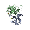
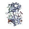
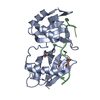
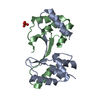
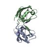
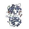
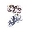
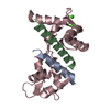
| Title | 2.1 Ang structure of the dimerized PAS domain of signal transduction histidine kinase from Nostoc punctiforme PCC 73102 with homology to the H-NOXA/H-NOBA domain of the soluble guanylyl cyclase | ||||||
 Components Components | signal transduction histidine kinase | ||||||
 Keywords Keywords | TRANSFERASE / PAS-like fold dimer / homologous to soluble guanylyl cyclase domain | ||||||
| Function / homology |  Function and homology information Function and homology information | ||||||
| Biological species |  Nostoc punctiforme (bacteria) Nostoc punctiforme (bacteria) | ||||||
| Method |  X-RAY DIFFRACTION / SYNCHROTRON / MOLECULAR REPLACEMENT / Resolution: 2.11 Å X-RAY DIFFRACTION / SYNCHROTRON / MOLECULAR REPLACEMENT / Resolution: 2.11 Å | ||||||
 Authors Authors | van den Akker, F. / Ma, X. | ||||||
 Citation Citation | Journal: J.Biol.Chem. / Year: 2008 Title: PAS-mediated dimerization of soluble guanylyl cyclase revealed by signal transduction histidine kinase domain crystal structure. Authors: Ma, X. / Sayed, N. / Baskaran, P. / Beuve, A. / van den Akker, F. | ||||||
| History |
|
- Structure visualization
| Structure viewer | Molecule: MolmilJmol/JSmol |
|---|
- Downloads & links
-Download
| PDBx/mmCIF format | 2p04.cif.gz | 54.5 KB | Display | PDBx/mmCIF format |
|---|---|---|---|---|
| PDB format | pdb2p04.ent.gz | 40.7 KB | Display | PDB format |
| PDBx/mmJSON format | 2p04.json.gz | Tree view | PDBx/mmJSON format | |
| Others |  Other downloads Other downloads |
-Validation report
| Arichive directory | https://data.pdbj.org/pub/pdb/validation_reports/p0/2p04ftp://data.pdbj.org/pub/pdb/validation_reports/p0/2p04 | HTTPS FTP |
|---|
-Related structure data


-Links
 PDBj
PDBj

- Assembly
| Deposited unit | 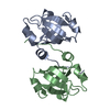
| ||||||||
|---|---|---|---|---|---|---|---|---|---|
| 1 |
| ||||||||
| Unit cell |
|
-Components
| #1: Protein | Mass: 13870.230 Da / Num. of mol.: 2 / Fragment: H-NOXA/H-NOBA domain Source method: isolated from a genetically manipulated source Source: (gene. exp.) Nostoc punctiforme (bacteria) / Strain: PCC 73102 / Gene: COG0642 / Plasmid: pET28a / Species (production host): Escherichia coli / Production host: References: UniProt: D0VWX5*PLUS, Transferases; Transferring phosphorus-containing groups; Phosphotransferases with a nitrogenous group as acceptor #2: Water | ChemComp-HOH / |  Mass: 18.015 Da / Num. of mol.: 55 / Source method: isolated from a natural source / Formula: H2O Mass: 18.015 Da / Num. of mol.: 55 / Source method: isolated from a natural source / Formula: H2O |
|---|
-Experimental details
-Experiment
| Experiment | Method: X-RAY DIFFRACTION / Number of used crystals: 1 |
|---|
- Sample preparation
| Crystal | Density Matthews: 2.2 Å3/Da / Density % sol: 44.08 % |
|---|---|
| Crystal grow | Temperature: 298 K / Method: vapor diffusion, sitting drop / pH: 7.5 Details: 0.1M Hepes pH 7.5, 1.5M LiSO4, VAPOR DIFFUSION, SITTING DROP, temperature 298K |
-Data collection
| Diffraction | Mean temperature: 100 K |
|---|---|
| Diffraction source | Source: SYNCHROTRON / Site: APS  / Beamline: 19-ID / Wavelength: 0.97934 / Beamline: 19-ID / Wavelength: 0.97934 |
| Detector | Type: ADSC QUANTUM 315 / Detector: CCD / Date: Jun 8, 2006 |
| Radiation | Protocol: SINGLE WAVELENGTH / Monochromatic (M) / Laue (L): M / Scattering type: x-ray |
| Radiation wavelength | Wavelength: 0.97934 Å / Relative weight: 1 |
| Reflection | Resolution: 2.1→50 Å / Num. obs: 12364 / % possible obs: 87.4 % / Observed criterion σ(I): 0 / Redundancy: 3.4 % / Rsym value: 0.119 / Net I/σ(I): 10 |
| Reflection shell | Resolution: 2.1→2.18 Å / Redundancy: 1.9 % / Mean I/σ(I) obs: 1.6 / Rsym value: 0.456 / % possible all: 59.8 |
- Processing
| Software |
| ||||||||||||||||||||||||||||||||||||||||||||||||||||||||||||||||||||||||||||||||||||||||||||||||||||||||||||||||||||||||||||||||||||||||||||||||||||||||||||||||||||||||||
|---|---|---|---|---|---|---|---|---|---|---|---|---|---|---|---|---|---|---|---|---|---|---|---|---|---|---|---|---|---|---|---|---|---|---|---|---|---|---|---|---|---|---|---|---|---|---|---|---|---|---|---|---|---|---|---|---|---|---|---|---|---|---|---|---|---|---|---|---|---|---|---|---|---|---|---|---|---|---|---|---|---|---|---|---|---|---|---|---|---|---|---|---|---|---|---|---|---|---|---|---|---|---|---|---|---|---|---|---|---|---|---|---|---|---|---|---|---|---|---|---|---|---|---|---|---|---|---|---|---|---|---|---|---|---|---|---|---|---|---|---|---|---|---|---|---|---|---|---|---|---|---|---|---|---|---|---|---|---|---|---|---|---|---|---|---|---|---|---|---|---|---|
| Refinement | Method to determine structure: MOLECULAR REPLACEMENT Starting model: Truncated delta-7 H-NOXA structure Resolution: 2.11→25.26 Å / Cor.coef. Fo:Fc: 0.951 / Cor.coef. Fo:Fc free: 0.918 / SU B: 6.062 / SU ML: 0.16 / Cross valid method: THROUGHOUT / ESU R: 0.282 / ESU R Free: 0.23 / Stereochemistry target values: MAXIMUM LIKELIHOOD
| ||||||||||||||||||||||||||||||||||||||||||||||||||||||||||||||||||||||||||||||||||||||||||||||||||||||||||||||||||||||||||||||||||||||||||||||||||||||||||||||||||||||||||
| Solvent computation | Ion probe radii: 0.8 Å / Shrinkage radii: 0.8 Å / VDW probe radii: 1.2 Å / Solvent model: MASK | ||||||||||||||||||||||||||||||||||||||||||||||||||||||||||||||||||||||||||||||||||||||||||||||||||||||||||||||||||||||||||||||||||||||||||||||||||||||||||||||||||||||||||
| Displacement parameters | Biso mean: 39.334 Å2
| ||||||||||||||||||||||||||||||||||||||||||||||||||||||||||||||||||||||||||||||||||||||||||||||||||||||||||||||||||||||||||||||||||||||||||||||||||||||||||||||||||||||||||
| Refinement step | Cycle: LAST / Resolution: 2.11→25.26 Å
| ||||||||||||||||||||||||||||||||||||||||||||||||||||||||||||||||||||||||||||||||||||||||||||||||||||||||||||||||||||||||||||||||||||||||||||||||||||||||||||||||||||||||||
| Refine LS restraints |
| ||||||||||||||||||||||||||||||||||||||||||||||||||||||||||||||||||||||||||||||||||||||||||||||||||||||||||||||||||||||||||||||||||||||||||||||||||||||||||||||||||||||||||
| LS refinement shell | Highest resolution: 2.11 Å / Num. reflection Rwork: 556 / Total num. of bins used: 20 |
