 ムービー
ムービー コントローラー


+ データを開く

- 基本情報
基本情報
| 登録情報 | データベース: PDB / ID: 2oya | ||||||
|---|---|---|---|---|---|---|---|
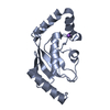
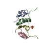
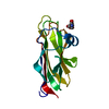
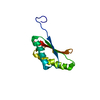
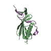
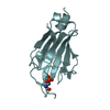
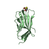
| タイトル | Crystal structure analysis of the dimeric form of the SRCR domain of mouse MARCO | ||||||
 要素 要素 | Macrophage receptor MARCO | ||||||
 キーワード キーワード | LIGAND BINDING PROTEIN / Extracellular matrix / Scavenger receptor cysteine-rich (SRCR) / macrophage receptor / ligand binding / basic cluster / acidic cluster / sulfate binding / dimer | ||||||
| 機能・相同性 |  機能・相同性情報 機能・相同性情報Scavenging by Class A Receptors / collagen trimer / apoptotic cell clearance / cargo receptor activity / phagocytosis, engulfment / amyloid-beta clearance / receptor-mediated endocytosis / G protein-coupled receptor binding / endocytosis / adenylate cyclase-inhibiting G protein-coupled receptor signaling pathway ...Scavenging by Class A Receptors / collagen trimer / apoptotic cell clearance / cargo receptor activity / phagocytosis, engulfment / amyloid-beta clearance / receptor-mediated endocytosis / G protein-coupled receptor binding / endocytosis / adenylate cyclase-inhibiting G protein-coupled receptor signaling pathway / amyloid-beta binding / positive regulation of ERK1 and ERK2 cascade / innate immune response / plasma membrane / cytoplasm 類似検索 - 分子機能 | ||||||
| 生物種 |  | ||||||
| 手法 |  X線回折 / シンクロトロン / 分子置換 / 解像度: 1.77 Å X線回折 / シンクロトロン / 分子置換 / 解像度: 1.77 Å | ||||||
 データ登録者 データ登録者 | Ojala, J.R.M. / Pikkarainen, T. / Tuuttila, A. / Sandalova, T. / Tryggvason, K. | ||||||
 引用 引用 | ジャーナル: J.Biol.Chem. / 年: 2007 タイトル: Crystal structure of the cysteine-rich domain of scavenger receptor MARCO reveals the presence of a basic and an acidic cluster that both contribute to ligand recognition. 著者: Ojala, J.R. / Pikkarainen, T. / Tuuttila, A. / Sandalova, T. / Tryggvason, K. | ||||||
| 履歴 |
|
- 構造の表示
構造の表示
| 構造ビューア | 分子: MolmilJmol/JSmol |
|---|
- ダウンロードとリンク
-ダウンロード
| PDBx/mmCIF形式 | 2oya.cif.gz | 57.9 KB | 表示 | PDBx/mmCIF形式 |
|---|---|---|---|---|
| PDB形式 | pdb2oya.ent.gz | 41.3 KB | 表示 | PDB形式 |
| PDBx/mmJSON形式 | 2oya.json.gz | ツリー表示 | PDBx/mmJSON形式 | |
| その他 |  その他のダウンロード その他のダウンロード |
-検証レポート
| アーカイブディレクトリ | https://data.pdbj.org/pub/pdb/validation_reports/oy/2oyaftp://data.pdbj.org/pub/pdb/validation_reports/oy/2oya | HTTPS FTP |
|---|
-関連構造データ


-リンク
 PDBj
PDBj- 集合体
集合体
| 登録構造単位 | 
| ||||||||
|---|---|---|---|---|---|---|---|---|---|
| 1 | 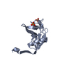
| ||||||||
| 2 | 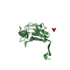
| ||||||||
| 3 |
| ||||||||
| 単位格子 |
| ||||||||
| 詳細 | Biological unit is a monomer. There are two biological units in the asymmetric unit (chains A & B) |
-要素
| #1: タンパク質 | 分子量: 11337.281 Da / 分子数: 2 断片: C-terminal domain, scavenger receptor cysteine-rich domain (SRCR) 由来タイプ: 組換発現 / 由来: (組換発現)  Homo sapiens (ヒト) / 参照: UniProt: Q60754 Homo sapiens (ヒト) / 参照: UniProt: Q60754#2: 化合物 |   分子量: 96.063 Da / 分子数: 3 / 由来タイプ: 合成 / 式: SO4 分子量: 96.063 Da / 分子数: 3 / 由来タイプ: 合成 / 式: SO4#3: 水 | ChemComp-HOH / |  分子量: 18.015 Da / 分子数: 192 / 由来タイプ: 天然 / 式: H2O 分子量: 18.015 Da / 分子数: 192 / 由来タイプ: 天然 / 式: H2OHas protein modification | Y | |
|---|
-実験情報
-実験
| 実験 | 手法: X線回折 / 使用した結晶の数: 1 |
|---|
- 試料調製
試料調製
| 結晶 | マシュー密度: 1.99 Å3/Da / 溶媒含有率: 38.11 % |
|---|---|
| 結晶化 | 温度: 277 K / 手法: 蒸気拡散法, ハンギングドロップ法 / pH: 6 詳細: 7% PEG 4000, 0.1M Bis-Tris, 0.1M Lithium sulfate, pH 6.0, VAPOR DIFFUSION, HANGING DROP, temperature 277K |
-データ収集
| 回折 | 平均測定温度: 100 K |
|---|---|
| 放射光源 | 由来: シンクロトロン / サイト: MAX II  / ビームライン: I711 / 波長: 1.097 Å / ビームライン: I711 / 波長: 1.097 Å |
| 検出器 | タイプ: MAR CCD 130 mm / 検出器: CCD / 日付: 2003年2月10日 |
| 放射 | プロトコル: SINGLE WAVELENGTH / 単色(M)・ラウエ(L): M / 散乱光タイプ: x-ray |
| 放射波長 | 波長: 1.097 Å / 相対比: 1 |
| 反射 | 解像度: 1.77→14.912 Å / Num. all: 17081 / Num. obs: 16364 / % possible obs: 95.4 % / Observed criterion σ(F): 0.013 / Observed criterion σ(I): 0 / 冗長度: 3.5 % / Rmerge(I) obs: 0.037 / Rsym value: 0.037 / Net I/σ(I): 13.8 |
| 反射 シェル | 解像度: 1.77→1.86 Å / 冗長度: 3.4 % / Rmerge(I) obs: 0.074 / Mean I/σ(I) obs: 9.1 / Num. measured all: 7835 / Num. unique all: 2282 / Rsym value: 0.074 / % possible all: 89.4 |
-位相決定
| Phasing MR | Rfactor: 0.428 / Cor.coef. Fo:Fc: 0.519
|
|---|
- 解析
解析
| ソフトウェア |
| ||||||||||||||||||||||||||||||||||||||||||||||||||||||||||||||||||||||||||||||||||||||||||
|---|---|---|---|---|---|---|---|---|---|---|---|---|---|---|---|---|---|---|---|---|---|---|---|---|---|---|---|---|---|---|---|---|---|---|---|---|---|---|---|---|---|---|---|---|---|---|---|---|---|---|---|---|---|---|---|---|---|---|---|---|---|---|---|---|---|---|---|---|---|---|---|---|---|---|---|---|---|---|---|---|---|---|---|---|---|---|---|---|---|---|---|
| 精密化 | 構造決定の手法: 分子置換 開始モデル: PDB ENTRY 2OY3 解像度: 1.77→14.912 Å / Cor.coef. Fo:Fc: 0.959 / Cor.coef. Fo:Fc free: 0.94 / SU B: 2.108 / SU ML: 0.069 / Isotropic thermal model: isotropic / 交差検証法: THROUGHOUT / σ(F): 0 / ESU R: 0.14 / ESU R Free: 0.126 / 立体化学のターゲット値: MAXIMUM LIKELIHOOD / 詳細: HYDROGENS HAVE BEEN ADDED IN THE RIDING POSITIONS
| ||||||||||||||||||||||||||||||||||||||||||||||||||||||||||||||||||||||||||||||||||||||||||
| 溶媒の処理 | イオンプローブ半径: 0.8 Å / 減衰半径: 0.8 Å / VDWプローブ半径: 1.4 Å / 溶媒モデル: MASK | ||||||||||||||||||||||||||||||||||||||||||||||||||||||||||||||||||||||||||||||||||||||||||
| 原子変位パラメータ | Biso mean: 11.59 Å2
| ||||||||||||||||||||||||||||||||||||||||||||||||||||||||||||||||||||||||||||||||||||||||||
| 精密化ステップ | サイクル: LAST / 解像度: 1.77→14.912 Å
| ||||||||||||||||||||||||||||||||||||||||||||||||||||||||||||||||||||||||||||||||||||||||||
| 拘束条件 |
| ||||||||||||||||||||||||||||||||||||||||||||||||||||||||||||||||||||||||||||||||||||||||||
| LS精密化 シェル | 解像度: 1.77→1.815 Å / Total num. of bins used: 20
|
