 Movie
Movie Controller

[English] 日本語
 Yorodumi
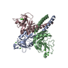
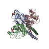
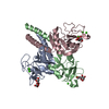
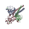
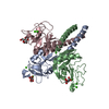
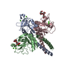
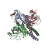
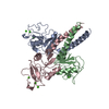
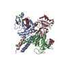
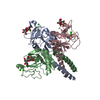
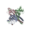
- PDB-2ork: crystal structure of the trimeric neck and carbohydrate recogniti... -
+ Open data

- Basic information
| Entry | Database: PDB / ID: 2ork | ||||||
|---|---|---|---|---|---|---|---|
| Title | crystal structure of the trimeric neck and carbohydrate recognition domain of human surfactant protein D in complex with inositol-1-phosphate | ||||||
 Components Components | Pulmonary surfactant-associated protein D | ||||||
 Keywords Keywords | SUGAR BINDING PROTEIN / Surfactant protein / Carbohydrate recognition domain / trimeric | ||||||
| Function / homology |  Function and homology information Function and homology informationToll Like Receptor TLR1:TLR2 Cascade / Defective CSF2RB causes SMDP5 / Defective CSF2RA causes SMDP4 / Toll Like Receptor 4 (TLR4) Cascade / clathrin-coated endocytic vesicle / respiratory gaseous exchange by respiratory system / Regulation of TLR by endogenous ligand / collagen trimer / surfactant homeostasis / Surfactant metabolism ...Toll Like Receptor TLR1:TLR2 Cascade / Defective CSF2RB causes SMDP5 / Defective CSF2RA causes SMDP4 / Toll Like Receptor 4 (TLR4) Cascade / clathrin-coated endocytic vesicle / respiratory gaseous exchange by respiratory system / Regulation of TLR by endogenous ligand / collagen trimer / surfactant homeostasis / Surfactant metabolism / Signal regulatory protein family interactions / negative regulation of interleukin-2 production / lung alveolus development / macrophage chemotaxis / endocytic vesicle / negative regulation of T cell proliferation / multivesicular body / regulation of cytokine production / reactive oxygen species metabolic process / positive regulation of phagocytosis / receptor-mediated endocytosis / Immunoregulatory interactions between a Lymphoid and a non-Lymphoid cell / SARS-CoV-1 activates/modulates innate immune responses / carbohydrate binding / lysosome / defense response to bacterium / innate immune response / endoplasmic reticulum membrane / SARS-CoV-2 activates/modulates innate and adaptive immune responses / : / extracellular region / identical protein binding Similarity search - Function | ||||||
| Biological species |  Homo sapiens (human) Homo sapiens (human) | ||||||
| Method |  X-RAY DIFFRACTION / SYNCHROTRON / FOURIER SYNTHESIS / Resolution: 1.89 Å X-RAY DIFFRACTION / SYNCHROTRON / FOURIER SYNTHESIS / Resolution: 1.89 Å | ||||||
 Authors Authors | Head, J.F. | ||||||
 Citation Citation | Journal: Biochemistry / Year: 2007 Title: Critical Role of Arg/Lys343 in the Species-Dependent Recognition of Phosphatidylinositol by Pulmonary Surfactant Protein D. Authors: Crouch, E. / McDonald, B. / Smith, K. / Roberts, M. / Mealy, T. / Seaton, B. / Head, J. | ||||||
| History |
|
- Structure visualization
| Structure viewer | Molecule: MolmilJmol/JSmol |
|---|
- Downloads & links
-Download
| PDBx/mmCIF format | 2ork.cif.gz | 115.2 KB | Display | PDBx/mmCIF format |
|---|---|---|---|---|
| PDB format | pdb2ork.ent.gz | 86.8 KB | Display | PDB format |
| PDBx/mmJSON format | 2ork.json.gz | Tree view | PDBx/mmJSON format | |
| Others |  Other downloads Other downloads |
-Validation report
| Arichive directory | https://data.pdbj.org/pub/pdb/validation_reports/or/2orkftp://data.pdbj.org/pub/pdb/validation_reports/or/2ork | HTTPS FTP |
|---|
-Related structure data
| Related structure data |  2orjC  2os9C 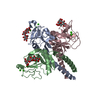 2gguS S: Starting model for refinement C: citing same article ( |
|---|---|
| Similar structure data |


-Links
 PDBj
PDBj


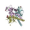
- Assembly
| Deposited unit | 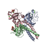
| ||||||||
|---|---|---|---|---|---|---|---|---|---|
| 1 |
| ||||||||
| Unit cell |
| ||||||||
| Details | The single trimer of head and neck domains present in the asymmetric unit is the functional unit. |
-Components
| #1: Protein | Mass: 17272.209 Da / Num. of mol.: 3 / Fragment: head and neck domain Source method: isolated from a genetically manipulated source Source: (gene. exp.) Homo sapiens (human) / Gene: SFTPD, PSPD, SFTP4 / Production host:  #2: Chemical | ChemComp-CA /   Mass: 40.078 Da / Num. of mol.: 9 / Source method: obtained synthetically / Formula: Ca Mass: 40.078 Da / Num. of mol.: 9 / Source method: obtained synthetically / Formula: Ca#3: Chemical | 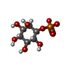  Mass: 258.120 Da / Num. of mol.: 2 / Source method: obtained synthetically / Formula: C6H11O9P Mass: 258.120 Da / Num. of mol.: 2 / Source method: obtained synthetically / Formula: C6H11O9P#4: Water | ChemComp-HOH / |  Mass: 18.015 Da / Num. of mol.: 517 / Source method: isolated from a natural source / Formula: H2O Mass: 18.015 Da / Num. of mol.: 517 / Source method: isolated from a natural source / Formula: H2OHas protein modification | Y | |
|---|
-Experimental details
-Experiment
| Experiment | Method: X-RAY DIFFRACTION / Number of used crystals: 1 |
|---|
- Sample preparation
| Crystal | Density Matthews: 3.21 Å3/Da / Density % sol: 61.72 % |
|---|---|
| Crystal grow | Temperature: 290 K / Method: vapor diffusion, hanging drop / pH: 7.5 Details: 150mM NaCl, 10mM CaCl2, 12% PEG 8000, 100mM HEPES , pH 7.5, VAPOR DIFFUSION, HANGING DROP, temperature 290K |
-Data collection
| Diffraction | Mean temperature: 100 K |
|---|---|
| Diffraction source | Source: SYNCHROTRON / Site: NSLS  / Beamline: X8C / Wavelength: 1 Å / Beamline: X8C / Wavelength: 1 Å |
| Detector | Type: ADSC QUANTUM 4 / Detector: CCD / Date: Jul 29, 2006 |
| Radiation | Monochromator: Si 111 double crystal / Protocol: SINGLE WAVELENGTH / Monochromatic (M) / Laue (L): M / Scattering type: x-ray |
| Radiation wavelength | Wavelength: 1 Å / Relative weight: 1 |
| Reflection | Resolution: 1.9→38.66 Å / Num. all: 51917 / Num. obs: 48123 / % possible obs: 92.7 % / Observed criterion σ(F): 0 / Observed criterion σ(I): 0 / Redundancy: 3.8 % / Rmerge(I) obs: 0.038 / Rsym value: 0.038 / Net I/σ(I): 46.2 |
| Reflection shell | Resolution: 1.9→1.97 Å / Redundancy: 2 % / Rmerge(I) obs: 0.067 / Mean I/σ(I) obs: 17 / Num. unique all: 3737 / Rsym value: 0.067 / % possible all: 72.6 |
- Processing
| Software |
| |||||||||||||||||||||||||||||||||||||||||||||||||||||||||||||||||||||||||||||||||||||||||||||||
|---|---|---|---|---|---|---|---|---|---|---|---|---|---|---|---|---|---|---|---|---|---|---|---|---|---|---|---|---|---|---|---|---|---|---|---|---|---|---|---|---|---|---|---|---|---|---|---|---|---|---|---|---|---|---|---|---|---|---|---|---|---|---|---|---|---|---|---|---|---|---|---|---|---|---|---|---|---|---|---|---|---|---|---|---|---|---|---|---|---|---|---|---|---|---|---|---|
| Refinement | Method to determine structure: FOURIER SYNTHESIS Starting model: PDB entry 2GGU Resolution: 1.89→38.66 Å / Cor.coef. Fo:Fc: 0.931 / Cor.coef. Fo:Fc free: 0.897 / SU B: 2.402 / SU ML: 0.074 / Cross valid method: THROUGHOUT / σ(F): 0 / σ(I): 0 / ESU R: 0.141 / ESU R Free: 0.136 / Stereochemistry target values: MAXIMUM LIKELIHOOD / Details: HYDROGENS HAVE BEEN ADDED IN THE RIDING POSITIONS
| |||||||||||||||||||||||||||||||||||||||||||||||||||||||||||||||||||||||||||||||||||||||||||||||
| Solvent computation | Ion probe radii: 0.8 Å / Shrinkage radii: 0.8 Å / VDW probe radii: 1.4 Å / Solvent model: MASK | |||||||||||||||||||||||||||||||||||||||||||||||||||||||||||||||||||||||||||||||||||||||||||||||
| Displacement parameters | Biso mean: 21.249 Å2
| |||||||||||||||||||||||||||||||||||||||||||||||||||||||||||||||||||||||||||||||||||||||||||||||
| Refinement step | Cycle: LAST / Resolution: 1.89→38.66 Å
| |||||||||||||||||||||||||||||||||||||||||||||||||||||||||||||||||||||||||||||||||||||||||||||||
| Refine LS restraints |
| |||||||||||||||||||||||||||||||||||||||||||||||||||||||||||||||||||||||||||||||||||||||||||||||
| LS refinement shell | Resolution: 1.9→1.94 Å / Total num. of bins used: 20
|
