[English] 日本語
Yorodumi
- PDB-2oqm: Crystal structure of a dinb family member protein (sden_0562) fro... -

+
Open data


ID or keywords:

Loading...

-
Basic information

Entry
Database: PDB / ID: 2oqm
Title
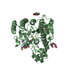
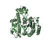
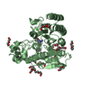
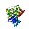
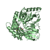
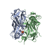
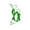
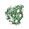
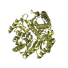
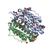
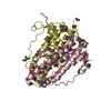
Crystal structure of a dinb family member protein (sden_0562) from shewanella denitrificans at 1.83 A resolution
ComponentsHypothetical protein
KeywordsUNKNOWN FUNCTION / Structural genomics / Joint Center for Structural Genomics / JCSG / Protein Structure Initiative / PSI-2
Function / homologyProtein of unknown function DUF1993 / Domain of unknown function (DUF1993) / dinb family like domain / DinB/YfiT-like putative metalloenzymes / Four Helix Bundle (Hemerythrin (Met), subunit A) / Up-down Bundle / Mainly Alpha / FORMIC ACID / Uncharacterized protein
Function and homology information
Biological speciesShewanella denitrificans (bacteria)
MethodX-RAY DIFFRACTION / SYNCHROTRON / MAD / Resolution: 1.83 Å
AuthorsJoint Center for Structural Genomics (JCSG)
CitationJournal: To be published
Title: Crystal structure of hypothetical protein (YP_561575.1) from Shewanella denitrificans OS-217 at 1.83 A resolution
Authors: Joint Center for Structural Genomics (JCSG)
History
DepositionJan 31, 2007Deposition site: RCSB / Processing site: RCSB
Revision 1.0Feb 13, 2007Provider: repository / Type: Initial release
Revision 1.1May 1, 2008Group: Version format compliance
Revision 1.2Jul 13, 2011Group: Advisory / Version format compliance
Revision 1.3Oct 18, 2017Group: Refinement description / Category: software / Item: _software.classification / _software.name
Revision 1.4Jan 25, 2023Group: Database references / Derived calculations
Category: database_2 / struct_conn ...database_2 / struct_conn / struct_ref_seq_dif / struct_site
Item: _database_2.pdbx_DOI / _database_2.pdbx_database_accession ..._database_2.pdbx_DOI / _database_2.pdbx_database_accession / _struct_conn.pdbx_leaving_atom_flag / _struct_ref_seq_dif.details / _struct_site.pdbx_auth_asym_id / _struct_site.pdbx_auth_comp_id / _struct_site.pdbx_auth_seq_id
Remark 999 SEQUENCE THE CONSTRUCT WAS EXPRESSED WITH A PURIFICATION TAG MGSDKIHHHHHHENLYFQG.

-
Structure visualization

Structure viewerMolecule:
MolmilJmol/JSmol

Downloads & links

-
Assembly

Deposited unit
A: Hypothetical protein
B: Hypothetical protein
C: Hypothetical protein
D: Hypothetical protein
hetero molecules


Theoretical massNumber of molelcules
Total (without water)90,02046
Polymers87,8254
Non-polymers2,19542
Water9,818545
1
A: Hypothetical protein
B: Hypothetical protein
hetero molecules


Theoretical massNumber of molelcules
Total (without water)45,22427
Polymers43,9132
Non-polymers1,31125
Water362
TypeNameSymmetry operationNumber
identity operation1_555x,y,z1
Buried area7980 Å2
ΔGint-16 kcal/mol
Surface area15370 Å2
MethodPISA
2
C: Hypothetical protein
D: Hypothetical protein
hetero molecules


Theoretical massNumber of molelcules
Total (without water)44,79719
Polymers43,9132
Non-polymers88417
Water362
TypeNameSymmetry operationNumber
identity operation1_555x,y,z1
Buried area7980 Å2
ΔGint-20 kcal/mol
Surface area15660 Å2
MethodPISA
Unit cell
Length a, b, c (Å)62.437, 71.722, 93.489
Angle α, β, γ (deg.)90.000, 107.350, 90.000
Int Tables number4
Space group name H-MP1211
Noncrystallographic symmetry (NCS)NCS domain:
IDEns-IDDetails
11A
21B
31C
41D

NCS domain segments:

Component-ID: 1 / Ens-ID: 1 / Beg label comp-ID: GLY / End label comp-ID: TYR / Refine code: 4 / Auth seq-ID: 0 - 168 / Label seq-ID: 19 - 187

Dom-IDAuth asym-IDLabel asym-ID
1AA
2BB
3CC
4DD

-
Components

#1: Protein
Hypothetical protein


Mass: 21956.258 Da / Num. of mol.: 4
Source method: isolated from a genetically manipulated source
Source: (gene. exp.) Shewanella denitrificans (bacteria) / Strain: OS217, DSM 15013 / Gene: YP_561575.1, Sden_0562 / Plasmid: speedET / Production host: Escherichia coli (E. coli) / Strain (production host): HK100 / References: UniProt: Q12RS4
#2: Chemical
ChemComp-EDO / 1,2-ETHANEDIOL / ETHYLENE GLYCOL


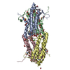
Mass: 62.068 Da / Num. of mol.: 17 / Source method: obtained synthetically / Formula: C2H6O2
#3: Chemical...
ChemComp-FMT / FORMIC ACID


Mass: 46.025 Da / Num. of mol.: 24 / Source method: obtained synthetically / Formula: CH2O2
#4: Chemical ChemComp-CL / CHLORIDE ION


Mass: 35.453 Da / Num. of mol.: 1 / Source method: obtained synthetically / Formula: Cl
#5: Water ChemComp-HOH / water


Mass: 18.015 Da / Num. of mol.: 545 / Source method: isolated from a natural source / Formula: H2O

-
Experimental details

-
Experiment

ExperimentMethod: X-RAY DIFFRACTION / Number of used crystals: 1

-
Sample preparation

CrystalDensity Matthews: 2.27 Å3/Da / Density % sol: 45.92 %
Crystal growTemperature: 293 K / Method: vapor diffusion, sitting drop
Details: NANODROP, 0.271M Ammonium formate, 18.5% Polyethylene glycol 3350, VAPOR DIFFUSION, SITTING DROP, temperature 293K

-
Data collection

DiffractionMean temperature: 100 K
Diffraction sourceSource: SYNCHROTRON / Site: SSRL / Beamline: BL11-1 / Wavelength: 0.91837, 0.97908, 0.97874
DetectorType: MARMOSAIC 325 mm CCD / Detector: CCD / Date: Jan 18, 2007 / Details: Flat mirror (vertical focusing)
RadiationMonochromator: Single crystal Si(111) bent (horizontal focusing)
Protocol: MAD / Monochromatic (M) / Laue (L): M / Scattering type: x-ray
Radiation wavelength
IDWavelength (Å)Relative weight
10.918371
20.979081
30.978741
ReflectionResolution: 1.83→45.22 Å / Num. obs: 68483 / % possible obs: 92.9 % / Biso Wilson estimate: 31.626 Å2 / Rmerge(I) obs: 0.065 / Net I/σ(I): 8.61
Reflection shell

Diffraction-ID: 1

Resolution (Å)Highest resolution (Å)Rmerge(I) obsMean I/σ(I) obsNum. measured obsNum. unique all% possible all
1.83-1.90.4672.214330615583.2
1.9-1.970.3732.712889552187.3
1.97-2.060.2823.514599624489.5
2.06-2.170.2114.615071647492.1
2.17-2.310.1575.915764676694.8
2.31-2.480.1227.214791632395.7
2.48-2.730.0889.215790677096.9
2.73-3.130.06612.116052686997
3.130.04216.715867675397.6

-
Phasing

PhasingMethod: MAD

-
Processing

Software
NameVersionClassificationNB
MolProbity3beta29model building
SHELXphasing
REFMAC5.2.0019refinement
XSCALEdata scaling
PDB_EXTRACT2data extraction
MAR345CCDdata collection
XDSdata reduction
SHELXDphasing
autoSHARPphasing
RefinementMethod to determine structure: MAD / Resolution: 1.83→45.22 Å / Cor.coef. Fo:Fc: 0.973 / Cor.coef. Fo:Fc free: 0.956 / SU B: 6.306 / SU ML: 0.093 / TLS residual ADP flag: LIKELY RESIDUAL / Cross valid method: THROUGHOUT / σ(F): 0 / ESU R: 0.118 / ESU R Free: 0.115
Stereochemistry target values: MAXIMUM LIKELIHOOD WITH PHASES
Details: 1. HYDROGENS HAVE BEEN ADDED IN THE RIDING POSITIONS. 2. ATOM RECORD CONTAINS RESIDUAL B FACTORS ONLY. 3. A MET-INHIBITION PROTOCOL WAS USED FOR SELENOMETHIONINE INCORPORATION DURING PROTEIN ...Details: 1. HYDROGENS HAVE BEEN ADDED IN THE RIDING POSITIONS. 2. ATOM RECORD CONTAINS RESIDUAL B FACTORS ONLY. 3. A MET-INHIBITION PROTOCOL WAS USED FOR SELENOMETHIONINE INCORPORATION DURING PROTEIN EXPRESSION. THE OCCUPANCY OF THE SE ATOMS IN THE MSE RESIDUES WAS REDUCED TO 0.75 FOR THE REDUCED SCATTERING POWER DUE TO PARTIAL S-MET INCORPORATION. 4. FMT, CL AND EDO ARE MODELED BASED ON THE CRYSTALLIZATION CONDITIONS OR CRYOPROTECTANT. 5. THE REGION D115-123 HAS POORLY DEFINED ELECTRON DENSITY.
RfactorNum. reflection% reflectionSelection details
Rfree0.196 3437 5 %RANDOM
Rwork0.157 ---
all0.159 ---
obs0.159 68463 98.4 %-
Solvent computationIon probe radii: 0.8 Å / Shrinkage radii: 0.8 Å / VDW probe radii: 1.2 Å / Solvent model: BABINET MODEL WITH MASK
Displacement parametersBiso mean: 24.558 Å2
Baniso -1Baniso -2Baniso -3
1--0.79 Å20 Å2-2.01 Å2
2---0.3 Å20 Å2
3----0.11 Å2
Refinement stepCycle: LAST / Resolution: 1.83→45.22 Å
ProteinNucleic acidLigandSolventTotal
Num. atoms5581 0 141 545 6267
Refine LS restraints
Refine-IDTypeDev idealDev ideal targetNumber
X-RAY DIFFRACTIONr_bond_refined_d0.0180.0225881
X-RAY DIFFRACTIONr_bond_other_d0.0080.023903
X-RAY DIFFRACTIONr_angle_refined_deg1.4921.9627921
X-RAY DIFFRACTIONr_angle_other_deg1.08939519
X-RAY DIFFRACTIONr_dihedral_angle_1_deg4.8675740
X-RAY DIFFRACTIONr_dihedral_angle_2_deg36.01524.61269
X-RAY DIFFRACTIONr_dihedral_angle_3_deg13.14115966
X-RAY DIFFRACTIONr_dihedral_angle_4_deg21.2311526
X-RAY DIFFRACTIONr_chiral_restr0.0940.2892
X-RAY DIFFRACTIONr_gen_planes_refined0.0070.026529
X-RAY DIFFRACTIONr_gen_planes_other0.0010.021189
X-RAY DIFFRACTIONr_nbd_refined0.2270.21366
X-RAY DIFFRACTIONr_nbd_other0.1880.24276
X-RAY DIFFRACTIONr_nbtor_refined0.1920.22890
X-RAY DIFFRACTIONr_nbtor_other0.0910.22853
X-RAY DIFFRACTIONr_xyhbond_nbd_refined0.1710.2429
X-RAY DIFFRACTIONr_symmetry_vdw_refined0.2560.219
X-RAY DIFFRACTIONr_symmetry_vdw_other0.2790.247
X-RAY DIFFRACTIONr_symmetry_hbond_refined0.1660.215
X-RAY DIFFRACTIONr_mcbond_it2.00233753
X-RAY DIFFRACTIONr_mcbond_other0.8431486
X-RAY DIFFRACTIONr_mcangle_it2.76155768
X-RAY DIFFRACTIONr_scbond_it4.85782419
X-RAY DIFFRACTIONr_scangle_it6.534112140
Refine LS restraints NCS

Ens-ID: 1 / Number: 2051 / Refine-ID: X-RAY DIFFRACTION

Dom-IDAuth asym-IDTypeRms dev position (Å)Weight position
1AMEDIUM POSITIONAL0.320.5
2BMEDIUM POSITIONAL0.40.5
3CMEDIUM POSITIONAL0.310.5
4DMEDIUM POSITIONAL0.570.5
1AMEDIUM THERMAL1.352
2BMEDIUM THERMAL1.382
3CMEDIUM THERMAL1.122
4DMEDIUM THERMAL1.322
LS refinement shellResolution: 1.83→1.88 Å / Total num. of bins used: 20
RfactorNum. reflection% reflection
Rfree0.278 246 -
Rwork0.261 4772 -
obs-5018 98.33 %
Refinement TLS params.

Method: refined / Refine-ID: X-RAY DIFFRACTION

IDL112)L122)L132)L222)L232)L332)S11 (Å °)S12 (Å °)S13 (Å °)S21 (Å °)S22 (Å °)S23 (Å °)S31 (Å °)S32 (Å °)S33 (Å °)T112)T122)T132)T222)T232)T332)Origin x (Å)Origin y (Å)Origin z (Å)
10.3954-0.25160.22410.7747-0.19521.12870.07270.0283-0.1394-0.0367-0.00860.13320.1707-0.0447-0.0642-0.0442-0.01370.0039-0.0641-0.01930.018916.5-13.17730.063
20.7948-0.06280.40430.6328-0.47081.3956-0.04070.14470.03250.0073-0.0653-0.0261-0.11650.22850.106-0.0441-0.02370.0273-0.01610.0005-0.034931.163-1.53724.514
30.7546-0.57330.49170.4562-0.55751.965-0.04350.10930.19550.0545-0.1077-0.1706-0.16730.57850.1512-0.0445-0.05520.00060.13350.01270.031251.92-10.1463.521
40.9775-0.34290.67080.4825-0.4641.5647-0.0231-0.16950.02820.11310.04740.0317-0.0827-0.0196-0.0243-0.01620.00140.04-0.022-0.0211-0.027834.472-12.97871.825
Refinement TLS group

Refine-ID: X-RAY DIFFRACTION / Selection: ALL

IDRefine TLS-IDAuth asym-IDLabel asym-IDAuth seq-IDLabel seq-ID
11AA-8 - 17111 - 190
22BB-4 - 17315 - 192
33CC-5 - 17314 - 192
44DD-6 - 17113 - 190

+
About Yorodumi

-
News

-
Feb 9, 2022. New format data for meta-information of EMDB entries

New format data for meta-information of EMDB entries

  • Version 3 of the EMDB header file is now the official format.
  • The previous official version 1.9 will be removed from the archive.

Related info.:EMDB header

External links:wwPDB to switch to version 3 of the EMDB data model

-
Aug 12, 2020. Covid-19 info

Covid-19 info

URL: https://pdbjlvh1.pdbj.org/emnavi/covid19.php

New page: Covid-19 featured information page in EM Navigator.

Related info.:Covid-19 info / Mar 5, 2020. Novel coronavirus structure data

+
Mar 5, 2020. Novel coronavirus structure data

Novel coronavirus structure data

Related info.:Yorodumi Speices / Aug 12, 2020. Covid-19 info

External links:COVID-19 featured content - PDBj / Molecule of the Month (242):Coronavirus Proteases

+
Jan 31, 2019. EMDB accession codes are about to change! (news from PDBe EMDB page)

EMDB accession codes are about to change! (news from PDBe EMDB page)

  • The allocation of 4 digits for EMDB accession codes will soon come to an end. Whilst these codes will remain in use, new EMDB accession codes will include an additional digit and will expand incrementally as the available range of codes is exhausted. The current 4-digit format prefixed with “EMD-” (i.e. EMD-XXXX) will advance to a 5-digit format (i.e. EMD-XXXXX), and so on. It is currently estimated that the 4-digit codes will be depleted around Spring 2019, at which point the 5-digit format will come into force.
  • The EM Navigator/Yorodumi systems omit the EMD- prefix.

Related info.:Q: What is EMD? / ID/Accession-code notation in Yorodumi/EM Navigator

External links:EMDB Accession Codes are Changing Soon! / Contact to PDBj

+
Jul 12, 2017. Major update of PDB

Major update of PDB

  • wwPDB released updated PDB data conforming to the new PDBx/mmCIF dictionary.
  • This is a major update changing the version number from 4 to 5, and with Remediation, in which all the entries are updated.
  • In this update, many items about electron microscopy experimental information are reorganized (e.g. em_software).
  • Now, EM Navigator and Yorodumi are based on the updated data.

External links:wwPDB Remediation / Enriched Model Files Conforming to OneDep Data Standards Now Available in the PDB FTP Archive

-
Yorodumi

Thousand views of thousand structures

  • Yorodumi is a browser for structure data from EMDB, PDB, SASBDB, etc.
  • This page is also the successor to EM Navigator detail page, and also detail information page/front-end page for Omokage search.
  • The word "yorodu" (or yorozu) is an old Japanese word meaning "ten thousand". "mi" (miru) is to see.

Related info.:EMDB / PDB / SASBDB / Comparison of 3 databanks / Yorodumi Search / Aug 31, 2016. New EM Navigator & Yorodumi / Yorodumi Papers / Jmol/JSmol / Function and homology information / Changes in new EM Navigator and Yorodumi

Read more