 Movie
Movie Controller

[English] 日本語
 Yorodumi
- PDB-2ooy: Crystal structure of the adenylate sensor from AMP-activated prot... -
+ Open data

- Basic information
| Entry | Database: PDB / ID: 2ooy | ||||||
|---|---|---|---|---|---|---|---|
| Title | Crystal structure of the adenylate sensor from AMP-activated protein kinase complexed with ATP | ||||||
 Components Components |
| ||||||
 Keywords Keywords | TRANSFERASE / AMPK / kinase / AMP | ||||||
| Function / homology |  Function and homology information Function and homology informationpositive regulation of flocculation / positive regulation of cell cycle switching, mitotic to meiotic cell cycle / AMPK inhibits chREBP transcriptional activation activity / Carnitine shuttle / : / Energy dependent regulation of mTOR by LKB1-AMPK / TP53 Regulates Metabolic Genes / Macroautophagy / mitotic spindle pole body / regulation of carbon utilization ...positive regulation of flocculation / positive regulation of cell cycle switching, mitotic to meiotic cell cycle / AMPK inhibits chREBP transcriptional activation activity / Carnitine shuttle / : / Energy dependent regulation of mTOR by LKB1-AMPK / TP53 Regulates Metabolic Genes / Macroautophagy / mitotic spindle pole body / regulation of carbon utilization / SREBP signaling pathway / CAMKK-AMPK signaling cascade / nucleotide-activated protein kinase complex / protein kinase regulator activity / regulation of glycolytic process / AMP binding / protein kinase activator activity / cellular response to glucose starvation / negative regulation of cytoplasmic translation / negative regulation of TORC1 signaling / positive regulation of gluconeogenesis / ADP binding / non-specific serine/threonine protein kinase / protein serine kinase activity / protein serine/threonine kinase activity / regulation of transcription by RNA polymerase II / protein kinase binding / signal transduction / positive regulation of transcription by RNA polymerase II / ATP binding / nucleus / plasma membrane / cytoplasm / cytosol Similarity search - Function | ||||||
| Biological species |  | ||||||
| Method |  X-RAY DIFFRACTION / SYNCHROTRON / MOLECULAR REPLACEMENT / Resolution: 2.88 Å X-RAY DIFFRACTION / SYNCHROTRON / MOLECULAR REPLACEMENT / Resolution: 2.88 Å | ||||||
 Authors Authors | Townley, R. / Shapiro, L. | ||||||
 Citation Citation | Journal: Science / Year: 2007 Title: Crystal structures of the adenylate sensor from fission yeast AMP-activated protein kinase. Authors: Townley, R. / Shapiro, L. | ||||||
| History |
| ||||||
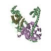
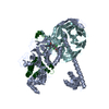
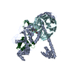
| Remark 300 | BIOMOLECULE: 1 THIS ENTRY CONTAINS THE CRYSTALLOGRAPHIC ASYMMETRIC UNIT WHICH CONSISTS OF 6 ... BIOMOLECULE: 1 THIS ENTRY CONTAINS THE CRYSTALLOGRAPHIC ASYMMETRIC UNIT WHICH CONSISTS OF 6 CHAIN(S). SEE REMARK 350 FOR INFORMATION ON GENERATING THE BIOLOGICAL MOLECULE(S). AUTHORS STATE THAT THE DEFINITIVE BIOLOGICAL UNIT IS A HETEROTRIMER (THERE ARE TWO SUCH TRIMERS: A+B+G AND C+D+E IN THE ASYMMETRIC UNIT), AND THAT THE DIMER OF THESE HETEROTRIMERS (SEE REMARK 350) IS ALSO PHYSIOLOGICALLY RELEVANT. |
- Structure visualization
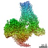
| Structure viewer | Molecule: MolmilJmol/JSmol |
|---|
- Downloads & links
-Download
| PDBx/mmCIF format | 2ooy.cif.gz | 238.4 KB | Display | PDBx/mmCIF format |
|---|---|---|---|---|
| PDB format | pdb2ooy.ent.gz | 186.8 KB | Display | PDB format |
| PDBx/mmJSON format | 2ooy.json.gz | Tree view | PDBx/mmJSON format | |
| Others |  Other downloads Other downloads |
-Validation report
| Arichive directory | https://data.pdbj.org/pub/pdb/validation_reports/oo/2ooyftp://data.pdbj.org/pub/pdb/validation_reports/oo/2ooy | HTTPS FTP |
|---|
-Related structure data
| Related structure data |  2ooxSC S: Starting model for refinement C: citing same article ( |
|---|---|
| Similar structure data |

-Links
 PDBj
PDBj


- Assembly
| Deposited unit | 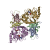
| ||||||||
|---|---|---|---|---|---|---|---|---|---|
| 1 |
| ||||||||
| Unit cell |
| ||||||||
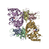
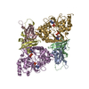
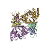
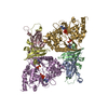
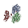
| Details | The biological assembly is a dimer in the asymmetric unit. |
-Components
-Protein , 3 types, 6 molecules ACBDGE
| #1: Protein | Mass: 15903.413 Da / Num. of mol.: 2 / Fragment: C-terminal domain: Residues 440-576 Source method: isolated from a genetically manipulated source Source: (gene. exp.) Strain: 972 / Gene: ssp2, SPCC74.03c / Plasmid: pSMT3, pET-Duet-1 / Species (production host): Escherichia coli / Production host:  References: UniProt: O74536, non-specific serine/threonine protein kinase #2: Protein | Mass: 10865.345 Da / Num. of mol.: 2 / Fragment: C-terminal domain: Residues 203-298 Source method: isolated from a genetically manipulated source Source: (gene. exp.) Strain: 972 / Gene: SPCC1919.03c / Plasmid: pSMT3, pET-Duet-1 / Species (production host): Escherichia coli / Production host: #3: Protein | Mass: 37364.992 Da / Num. of mol.: 2 Source method: isolated from a genetically manipulated source Source: (gene. exp.) Strain: 972 / Gene: SPAC1556.08c, SPAC1F12.01c / Plasmid: pSMT3, pET-Duet-1 / Species (production host): Escherichia coli / Production host: |
|---|
-Non-polymers , 3 types, 431 molecules 


| #4: Chemical |  Mass: 507.181 Da / Num. of mol.: 2 / Source method: obtained synthetically / Formula: C10H16N5O13P3 / Comment: ATP, energy-carrying molecule*YM Mass: 507.181 Da / Num. of mol.: 2 / Source method: obtained synthetically / Formula: C10H16N5O13P3 / Comment: ATP, energy-carrying molecule*YM#5: Chemical | ChemComp-FLC / |  Mass: 189.100 Da / Num. of mol.: 1 / Source method: obtained synthetically / Formula: C6H5O7 Mass: 189.100 Da / Num. of mol.: 1 / Source method: obtained synthetically / Formula: C6H5O7#6: Water | ChemComp-HOH / | Mass: 18.015 Da / Num. of mol.: 428 / Source method: isolated from a natural source / Formula: H2O |
|---|
-Experimental details
-Experiment
| Experiment | Method: X-RAY DIFFRACTION / Number of used crystals: 1 |
|---|
- Sample preparation
| Crystal | Density Matthews: 2.25 Å3/Da / Density % sol: 45.39 % |
|---|---|
| Crystal grow | Temperature: 293 K / Method: vapor diffusion, hanging drop / pH: 7.5 Details: 7.3-8.1% PEG3350, 0.1M HEPES, pH 7.5, 5mM ATP, VAPOR DIFFUSION, HANGING DROP, temperature 293K |
-Data collection
| Diffraction | Mean temperature: 100 K |
|---|---|
| Diffraction source | Source: SYNCHROTRON / Site: NSLS  / Beamline: X4A / Wavelength: 0.97898 Å / Beamline: X4A / Wavelength: 0.97898 Å |
| Detector | Type: ADSC QUANTUM 315 / Detector: CCD / Date: Jun 1, 2006 |
| Radiation | Monochromator: Si 111 CHANNEL / Protocol: SINGLE WAVELENGTH / Monochromatic (M) / Laue (L): M / Scattering type: x-ray |
| Radiation wavelength | Wavelength: 0.97898 Å / Relative weight: 1 |
| Reflection | Resolution: 2.88→50 Å / Num. all: 25963 / Num. obs: 24969 / % possible obs: 96.2 % / Observed criterion σ(I): 2 / Redundancy: 2.7 % / Rmerge(I) obs: 0.1 / Net I/σ(I): 8.3 |
| Reflection shell | Resolution: 2.88→3 Å / Rmerge(I) obs: 0.417 / % possible all: 93.4 |
- Processing
| Software |
| ||||||||||||||||||||||||||||||||||||||||||||||||||||||||||||||||||||||||||||||||||||||||||||||||||||||||||||||||||||||||||||||||||||||||||||||||||||||||||||||||||||||||||
|---|---|---|---|---|---|---|---|---|---|---|---|---|---|---|---|---|---|---|---|---|---|---|---|---|---|---|---|---|---|---|---|---|---|---|---|---|---|---|---|---|---|---|---|---|---|---|---|---|---|---|---|---|---|---|---|---|---|---|---|---|---|---|---|---|---|---|---|---|---|---|---|---|---|---|---|---|---|---|---|---|---|---|---|---|---|---|---|---|---|---|---|---|---|---|---|---|---|---|---|---|---|---|---|---|---|---|---|---|---|---|---|---|---|---|---|---|---|---|---|---|---|---|---|---|---|---|---|---|---|---|---|---|---|---|---|---|---|---|---|---|---|---|---|---|---|---|---|---|---|---|---|---|---|---|---|---|---|---|---|---|---|---|---|---|---|---|---|---|---|---|---|
| Refinement | Method to determine structure: MOLECULAR REPLACEMENT Starting model: PDB entry 2OOX Resolution: 2.88→50 Å / Cor.coef. Fo:Fc: 0.948 / Cor.coef. Fo:Fc free: 0.879 / SU B: 20.894 / SU ML: 0.4 / Cross valid method: THROUGHOUT / σ(F): 1 / ESU R Free: 0.546 / Stereochemistry target values: MAXIMUM LIKELIHOOD / Details: HYDROGENS HAVE BEEN ADDED IN THE RIDING POSITIONS
| ||||||||||||||||||||||||||||||||||||||||||||||||||||||||||||||||||||||||||||||||||||||||||||||||||||||||||||||||||||||||||||||||||||||||||||||||||||||||||||||||||||||||||
| Solvent computation | Ion probe radii: 0.8 Å / Shrinkage radii: 0.8 Å / VDW probe radii: 1.4 Å / Solvent model: MASK | ||||||||||||||||||||||||||||||||||||||||||||||||||||||||||||||||||||||||||||||||||||||||||||||||||||||||||||||||||||||||||||||||||||||||||||||||||||||||||||||||||||||||||
| Displacement parameters | Biso mean: 54.539 Å2
| ||||||||||||||||||||||||||||||||||||||||||||||||||||||||||||||||||||||||||||||||||||||||||||||||||||||||||||||||||||||||||||||||||||||||||||||||||||||||||||||||||||||||||
| Refinement step | Cycle: LAST / Resolution: 2.88→50 Å
| ||||||||||||||||||||||||||||||||||||||||||||||||||||||||||||||||||||||||||||||||||||||||||||||||||||||||||||||||||||||||||||||||||||||||||||||||||||||||||||||||||||||||||
| Refine LS restraints |
| ||||||||||||||||||||||||||||||||||||||||||||||||||||||||||||||||||||||||||||||||||||||||||||||||||||||||||||||||||||||||||||||||||||||||||||||||||||||||||||||||||||||||||
| LS refinement shell | Resolution: 2.88→2.96 Å / Total num. of bins used: 20
|
