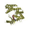
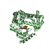
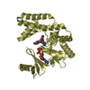
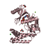
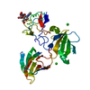
- PDB-2oh3: Crystal structure of COG1633: Uncharacterized conserved protein (... -
+
Open data
ID or keywords:
Loading...
-
Basic information
Entry
Database: PDB / ID: 2oh3
Title
Crystal structure of COG1633: Uncharacterized conserved protein (ZP_00055496.1) from Magnetospirillum magnetotacticum MS-1 at 2.00 A resolution
Components
COG1633: Uncharacterized conserved protein
Keywords
METAL BINDING PROTEIN / Rubrerythrin / ZP_00055496.1 / COG1633: Uncharacterized conserved protein / Structural Genomics / Joint Center for Structural Genomics / JCSG / Protein Structure Initiative / PSI-2
BIOMOLECULE: 1 THIS ENTRY CONTAINS THE CRYSTALLOGRAPHIC ASYMMETRIC UNIT WHICH CONSISTS OF 1 ... BIOMOLECULE: 1 THIS ENTRY CONTAINS THE CRYSTALLOGRAPHIC ASYMMETRIC UNIT WHICH CONSISTS OF 1 CHAIN(S). SEE REMARK 350 FOR INFORMATION ON GENERATING THE BIOLOGICAL MOLECULE(S). AUTHORS STATE THAT STATIC LIGHT SCATTERING WITH ANALYTICAL SIZE EXCLUSION CHROMATOGRAPHY MEASUREMENTS INDICATE THAT THE DIMER IS A BIOLOGICALLY SIGNIFICANT OLIGOMERIZATION STATE IN SOLUTION
Remark 999
SEQUENCE (1) THE CONSTRUCT WAS EXPRESSED WITH AN N-TERMINAL PURIFICATION TAG MGSDKIHHHHHHENLYFQG. ... SEQUENCE (1) THE CONSTRUCT WAS EXPRESSED WITH AN N-TERMINAL PURIFICATION TAG MGSDKIHHHHHHENLYFQG. THE TAG WAS REMOVED WITH TEV PROTEASE, LEAVING ONLY A GLYCINE (0), FOLLOWED BY THE TARGET SEQUENCE. (2) THE SEQUENCE OF THIS PROTEIN WAS NOT AVAILABLE AT THE UNIPROT DATABASE AT THE TIME OF DEPOSITION. (3) THE SEQUENCE IS AVAILABLE FROM GENBANK UNDER ACCESSION ID ZP_00055496.1 AND FROM THE UNIPROT ARCHIVE UNDER ACCESSION ID UPI00003846C6
Type: MARMOSAIC 325 mm CCD / Detector: CCD / Date: Dec 18, 2006 / Details: FLAT MIRROR (VERTICAL FOCUSING)
Radiation
Monochromator: SINGLE CRYSTAL SI(111) BENT (HORIZONTAL FOCUSING) Protocol: MAD / Monochromatic (M) / Laue (L): M / Scattering type: x-ray
Radiation wavelength
ID
Wavelength (Å)
Relative weight
1
0.91837
1
2
0.97913
1
Reflection
Resolution: 2→28.105 Å / Num. obs: 14778 / % possible obs: 96.8 % / Biso Wilson estimate: 37.83 Å2 / Rmerge(I) obs: 0.035 / Net I/σ(I): 16.21
Reflection shell
Resolution: 2→2.07 Å / Rmerge(I) obs: 0.365 / Mean I/σ(I) obs: 2.16 / % possible all: 94.5
-
Phasing
Phasing
Method: MAD
-
Processing
Software
Name
Version
Classification
NB
MolProbity
3beta29
modelbuilding
SHELX
phasing
REFMAC
5.2.0005
refinement
XSCALE
datascaling
PDB_EXTRACT
2
dataextraction
XDS
datareduction
SHELXD
phasing
autoSHARP
phasing
Refinement
Method to determine structure: MAD / Resolution: 2→28.11 Å / Cor.coef. Fo:Fc: 0.954 / Cor.coef. Fo:Fc free: 0.934 / SU B: 7.432 / SU ML: 0.111 / TLS residual ADP flag: LIKELY RESIDUAL / Cross valid method: THROUGHOUT / σ(F): 0 / ESU R: 0.156 / ESU R Free: 0.15 Stereochemistry target values: MAXIMUM LIKELIHOOD WITH PHASES Details: 1. HYDROGENS HAVE BEEN ADDED IN THE RIDING POSITIONS. 2. A MET-INHIBITION PROTOCOL WAS USED FOR SELENOMETHIONINE INCORPORATION DURING PROTEIN EXPRESSION. THE OCCUPANCY OF THE SE ATOMS IN THE ...Details: 1. HYDROGENS HAVE BEEN ADDED IN THE RIDING POSITIONS. 2. A MET-INHIBITION PROTOCOL WAS USED FOR SELENOMETHIONINE INCORPORATION DURING PROTEIN EXPRESSION. THE OCCUPANCY OF THE SE ATOMS IN THE MSE RESIDUES WAS REDUCED TO 0.75 FOR THE REDUCED SCATTERING POWER DUE TO PARTIAL S-MET INCORPORATION. 3. ATOM RECORD CONTAINS RESIDUAL B FACTORS ONLY. 4. THE RESIDUES 1-2, 85-87, AND 155-166 ARE DISORDERED AND WERE NOT BUILT DUE TO INSUFFICIENT ELECTRON DENSITY. 5. THE PRESENCE OF ZINC IS SUPPORTED WITH X-RAY FLOURESCENCE AND ANOMALOUS DIFFERENCE FOURIER MAPS. 6. IMIDAZOLE AND PEG WERE MODELED BASED ON CRYSTALLIZATION AND PURIFICATION CONDITIONS.
Rfactor
Num. reflection
% reflection
Selection details
Rfree
0.235
744
5 %
RANDOM
Rwork
0.192
-
-
-
obs
0.195
14778
97.7 %
-
Solvent computation
Ion probe radii: 0.8 Å / Shrinkage radii: 0.8 Å / VDW probe radii: 1.2 Å / Solvent model: MASK
Displacement parameters
Biso mean: 36.89 Å2
Baniso -1
Baniso -2
Baniso -3
1-
-2.24 Å2
0 Å2
0 Å2
2-
-
-0.27 Å2
0 Å2
3-
-
-
2.52 Å2
Refinement step
Cycle: LAST / Resolution: 2→28.11 Å
Protein
Nucleic acid
Ligand
Solvent
Total
Num. atoms
1178
0
26
111
1315
Refine LS restraints
Refine-ID
Type
Dev ideal
Dev ideal target
Number
X-RAY DIFFRACTION
r_bond_refined_d
0.019
0.022
1274
X-RAY DIFFRACTION
r_bond_other_d
0.003
0.02
1137
X-RAY DIFFRACTION
r_angle_refined_deg
1.491
1.962
1727
X-RAY DIFFRACTION
r_angle_other_deg
0.909
3
2644
X-RAY DIFFRACTION
r_dihedral_angle_1_deg
5.156
5
163
X-RAY DIFFRACTION
r_dihedral_angle_2_deg
39.194
23.968
63
X-RAY DIFFRACTION
r_dihedral_angle_3_deg
14.201
15
211
X-RAY DIFFRACTION
r_dihedral_angle_4_deg
22.839
15
9
X-RAY DIFFRACTION
r_chiral_restr
0.083
0.2
181
X-RAY DIFFRACTION
r_gen_planes_refined
0.006
0.02
1428
X-RAY DIFFRACTION
r_gen_planes_other
0.001
0.02
264
X-RAY DIFFRACTION
r_nbd_refined
0.225
0.2
290
X-RAY DIFFRACTION
r_nbd_other
0.193
0.2
1085
X-RAY DIFFRACTION
r_nbtor_refined
0.185
0.2
601
X-RAY DIFFRACTION
r_nbtor_other
0.089
0.2
720
X-RAY DIFFRACTION
r_xyhbond_nbd_refined
0.182
0.2
76
X-RAY DIFFRACTION
r_xyhbond_nbd_other
X-RAY DIFFRACTION
r_metal_ion_refined
0.092
0.2
3
X-RAY DIFFRACTION
r_metal_ion_other
X-RAY DIFFRACTION
r_symmetry_vdw_refined
0.283
0.2
12
X-RAY DIFFRACTION
r_symmetry_vdw_other
0.344
0.2
67
X-RAY DIFFRACTION
r_symmetry_hbond_refined
0.141
0.2
8
X-RAY DIFFRACTION
r_symmetry_hbond_other
X-RAY DIFFRACTION
r_symmetry_metal_ion_refined
X-RAY DIFFRACTION
r_symmetry_metal_ion_other
X-RAY DIFFRACTION
r_mcbond_it
0.904
1.5
786
X-RAY DIFFRACTION
r_mcbond_other
0.236
1.5
305
X-RAY DIFFRACTION
r_mcangle_it
1.479
2
1227
X-RAY DIFFRACTION
r_scbond_it
2.434
3
555
X-RAY DIFFRACTION
r_scangle_it
3.785
4.5
492
X-RAY DIFFRACTION
r_rigid_bond_restr
X-RAY DIFFRACTION
r_sphericity_free
X-RAY DIFFRACTION
r_sphericity_bonded
LS refinement shell
Resolution: 2→2.05 Å / Total num. of bins used: 20
Rfactor
Num. reflection
% reflection
Rfree
0.24
50
-
Rwork
0.25
1005
-
obs
-
-
95.22 %
Refinement TLS params.
Method: refined / Origin x: 31.0082 Å / Origin y: 24.553 Å / Origin z: 13.7822 Å
11
12
13
21
22
23
31
32
33
T
-0.1218 Å2
-0.0219 Å2
-0.0042 Å2
-
-0.1607 Å2
0.0345 Å2
-
-
-0.1768 Å2
L
5.1804 °2
-0.7722 °2
1.1 °2
-
1.7399 °2
-0.3541 °2
-
-
1.628 °2
S
-0.0297 Å °
0.2462 Å °
0.2082 Å °
-0.132 Å °
0.0244 Å °
0.0003 Å °
0.0029 Å °
0.0489 Å °
0.0053 Å °
+
About Yorodumi
-
News
-
Feb 9, 2022. New format data for meta-information of EMDB entries
New format data for meta-information of EMDB entries
Version 3 of the EMDB header file is now the official format.
The previous official version 1.9 will be removed from the archive.
In the structure databanks used in Yorodumi, some data are registered as the other names, "COVID-19 virus" and "2019-nCoV". Here are the details of the virus and the list of structure data.
Jan 31, 2019. EMDB accession codes are about to change! (news from PDBe EMDB page)
EMDB accession codes are about to change! (news from PDBe EMDB page)
The allocation of 4 digits for EMDB accession codes will soon come to an end. Whilst these codes will remain in use, new EMDB accession codes will include an additional digit and will expand incrementally as the available range of codes is exhausted. The current 4-digit format prefixed with “EMD-” (i.e. EMD-XXXX) will advance to a 5-digit format (i.e. EMD-XXXXX), and so on. It is currently estimated that the 4-digit codes will be depleted around Spring 2019, at which point the 5-digit format will come into force.
The EM Navigator/Yorodumi systems omit the EMD- prefix.
Related info.:Q: What is EMD? / ID/Accession-code notation in Yorodumi/EM Navigator
Yorodumi is a browser for structure data from EMDB, PDB, SASBDB, etc.
This page is also the successor to EM Navigator detail page, and also detail information page/front-end page for Omokage search.
The word "yorodu" (or yorozu) is an old Japanese word meaning "ten thousand". "mi" (miru) is to see.
Related info.:EMDB / PDB / SASBDB / Comparison of 3 databanks / Yorodumi Search / Aug 31, 2016. New EM Navigator & Yorodumi / Yorodumi Papers / Jmol/JSmol / Function and homology information / Changes in new EM Navigator and Yorodumi
 Movie
Movie Controller

 Yorodumi
 Open data
 Basic information
 Components
 Keywords
 Function and homology information
 Magnetospirillum magnetotacticum (bacteria)
 X-RAY DIFFRACTION /
  Authors
 Citation
 Structure visualization
 Downloads & links
 Other downloads
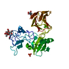


 PDBj
PDBj


 Assembly


 Mass: 65.409 Da / Num. of mol.: 1 / Source method: obtained synthetically / Formula: Zn
 Mass: 69.085 Da / Num. of mol.: 1 / Source method: obtained synthetically / Formula: C3H5N2
 Mass: 150.173 Da / Num. of mol.: 2 / Source method: obtained synthetically / Formula: C6H14O4
 Mass: 18.015 Da / Num. of mol.: 111 / Source method: isolated from a natural source / Formula: H2O
 Sample preparation
 / Beamline: BL11-1 / Wavelength: 0.91837, 0.97913
 Processing