 Movie
Movie Controller

[English] 日本語
 Yorodumi
- PDB-2ogk: Crystal structure of protein AF2318 from Archaeglobus fulgidus, P... -
+ Open data

- Basic information
| Entry | Database: PDB / ID: 2ogk | ||||||
|---|---|---|---|---|---|---|---|
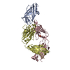
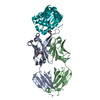
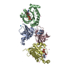
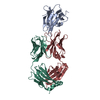
| Title | Crystal structure of protein AF2318 from Archaeglobus fulgidus, Pfam DUF54 | ||||||
 Components Components | Hypothetical protein | ||||||
 Keywords Keywords | STRUCTURAL GENOMICS / UNKNOWN FUNCTION / conserved hypothetical protein / Archaeglobus fulgidus / 10077d / PSI-2 / Protein Structure Initiative / New York SGX Research Center for Structural Genomics / NYSGXRC | ||||||
| Function / homology | Uncharacterised protein family UPF0201 / RNA binding / 50s Ribosomal Protein L5; Chain: A, / Ribosomal protein L5 / Ribosomal protein L5 domain superfamily / 2-Layer Sandwich / Alpha Beta / Exosome subunit Function and homology information Function and homology information | ||||||
| Biological species |   Archaeoglobus fulgidus (archaea) Archaeoglobus fulgidus (archaea) | ||||||
| Method |  X-RAY DIFFRACTION / SYNCHROTRON / SAD / Resolution: 3 Å X-RAY DIFFRACTION / SYNCHROTRON / SAD / Resolution: 3 Å | ||||||
 Authors Authors | Rao, K.N. / Burley, S.K. / Swaminathan, S. / New York SGX Research Center for Structural Genomics (NYSGXRC) | ||||||
 Citation Citation | Journal: Plos One / Year: 2008 Title: UPF201 archaeal specific family members reveal structural similarity to RNA-binding proteins but low likelihood for RNA-binding function. Authors: Rao, K.N. / Burley, S.K. / Swaminathan, S. | ||||||
| History |
|
- Structure visualization
| Structure viewer | Molecule: MolmilJmol/JSmol |
|---|
- Downloads & links
-Download
| PDBx/mmCIF format | 2ogk.cif.gz | 115.5 KB | Display | PDBx/mmCIF format |
|---|---|---|---|---|
| PDB format | pdb2ogk.ent.gz | 91 KB | Display | PDB format |
| PDBx/mmJSON format | 2ogk.json.gz | Tree view | PDBx/mmJSON format | |
| Others |  Other downloads Other downloads |
-Validation report
| Summary document | 2ogk_validation.pdf.gz | 446.6 KB | Display | wwPDB validaton report |
|---|---|---|---|---|
| Full document | 2ogk_full_validation.pdf.gz | 465.9 KB | Display | |
| Data in XML | 2ogk_validation.xml.gz | 23.5 KB | Display | |
| Data in CIF | 2ogk_validation.cif.gz | 31.5 KB | Display | |
| Arichive directory | https://data.pdbj.org/pub/pdb/validation_reports/og/2ogkftp://data.pdbj.org/pub/pdb/validation_reports/og/2ogk | HTTPS FTP |
-Related structure data
| Related structure data | 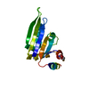 2nrqC  2nwuC 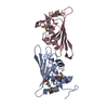 2pzzC C: citing same article ( |
|---|---|
| Similar structure data | |
| Other databases |

-Links
 PDBj
PDBj- Assembly
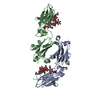
| Deposited unit | 
| ||||||||
|---|---|---|---|---|---|---|---|---|---|
| 1 |
| ||||||||
| Unit cell |
|
-Components
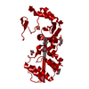
| #1: Protein | Mass: 16632.053 Da / Num. of mol.: 4 Source method: isolated from a genetically manipulated source Source: (gene. exp.) Archaeoglobus fulgidus (archaea) / Gene: AF_2318 / Production host:  #2: Water | ChemComp-HOH / |  Mass: 18.015 Da / Num. of mol.: 44 / Source method: isolated from a natural source / Formula: H2O Mass: 18.015 Da / Num. of mol.: 44 / Source method: isolated from a natural source / Formula: H2O |
|---|
-Experimental details
-Experiment
| Experiment | Method: X-RAY DIFFRACTION / Number of used crystals: 2 |
|---|
- Sample preparation
| Crystal | Density Matthews: 3.94 Å3/Da / Density % sol: 68.81 % |
|---|---|
| Crystal grow | Temperature: 293 K / Method: vapor diffusion, sitting drop / pH: 7 Details: PEG MME 5000, HEPES Buffer, Tacsimate, pH 7.0, VAPOR DIFFUSION, SITTING DROP, temperature 293K |
-Data collection
| Diffraction |
| ||||||||||||||||||
|---|---|---|---|---|---|---|---|---|---|---|---|---|---|---|---|---|---|---|---|
| Diffraction source |
| ||||||||||||||||||
| Detector |
| ||||||||||||||||||
| Radiation |
| ||||||||||||||||||
| Radiation wavelength |
| ||||||||||||||||||
| Reflection | Resolution: 3→50 Å / Num. all: 21971 / Num. obs: 21971 / % possible obs: 99.5 % / Observed criterion σ(F): 0 / Observed criterion σ(I): 0 / Redundancy: 6.6 % / Biso Wilson estimate: 40 Å2 / Rmerge(I) obs: 0.084 / Net I/σ(I): 8.7 | ||||||||||||||||||
| Reflection shell | Resolution: 3→3.12 Å / Redundancy: 6.1 % / Rmerge(I) obs: 0.446 / Mean I/σ(I) obs: 2.3 / Num. unique all: 2350 / % possible all: 99.1 |

- Processing
| Software |
| ||||||||||||||||||||||||||||||||||||
|---|---|---|---|---|---|---|---|---|---|---|---|---|---|---|---|---|---|---|---|---|---|---|---|---|---|---|---|---|---|---|---|---|---|---|---|---|---|
| Refinement | Method to determine structure: SAD / Resolution: 3→46.7 Å / Rfactor Rfree error: 0.015 / Data cutoff high absF: 55130.26 / Data cutoff low absF: 0 / Isotropic thermal model: RESTRAINED / Cross valid method: THROUGHOUT / σ(F): 0 / Stereochemistry target values: Engh & Huber Details: Residues listed as missing are due to lack of electron density, and residues for which side chain atoms are missing have been modeled as alanines
| ||||||||||||||||||||||||||||||||||||
| Solvent computation | Solvent model: FLAT MODEL / Bsol: 37.0738 Å2 / ksol: 0.311229 e/Å3 | ||||||||||||||||||||||||||||||||||||
| Displacement parameters | Biso mean: 63.3 Å2
| ||||||||||||||||||||||||||||||||||||
| Refine analyze |
| ||||||||||||||||||||||||||||||||||||
| Refinement step | Cycle: LAST / Resolution: 3→46.7 Å
| ||||||||||||||||||||||||||||||||||||
| Refine LS restraints |
| ||||||||||||||||||||||||||||||||||||
| LS refinement shell | Resolution: 3→3.19 Å / Rfactor Rfree error: 0.047 / Total num. of bins used: 6
| ||||||||||||||||||||||||||||||||||||
| Xplor file |
|
