 Movie
Movie Controller


+ Open data

- Basic information
| Entry | Database: PDB / ID: 2oep | ||||||
|---|---|---|---|---|---|---|---|
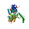
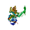
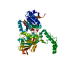
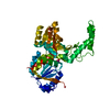
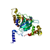
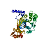
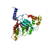
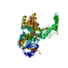
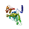
| Title | MSrecA-ADP-complex | ||||||
 Components Components | Protein recA | ||||||
 Keywords Keywords | RECOMBINATION / DNA-REPAIR / SOS RESPONCE | ||||||
| Function / homology |  Function and homology information Function and homology informationSOS response / ATP-dependent DNA damage sensor activity / single-stranded DNA binding / DNA recombination / damaged DNA binding / DNA repair / ATP binding / metal ion binding / cytosol Similarity search - Function | ||||||
| Biological species |  Mycobacterium smegmatis (bacteria) Mycobacterium smegmatis (bacteria) | ||||||
| Method |  X-RAY DIFFRACTION / MOLECULAR REPLACEMENT / Resolution: 3.1 Å X-RAY DIFFRACTION / MOLECULAR REPLACEMENT / Resolution: 3.1 Å | ||||||
 Authors Authors | Krishna, R. / Rajan Prabu, J. / Manjunath, G.P. / Datta, S. / Chandra, N.R. / Muniyappa, K. / Vijayan, M. | ||||||
 Citation Citation | Journal: J.Mol.Biol. / Year: 2007 Title: Snapshots of RecA protein involving movement of the C-domain and different conformations of the DNA-binding loops: crystallographic and comparative analysis of 11 structures of Mycobacterium smegmatis RecA Authors: Krishna, R. / Prabu, J.R. / Manjunath, G.P. / Datta, S. / Chandra, N.R. / Muniyappa, K. / Vijayan, M. #1: Journal: Nucleic Acids Res. / Year: 2000Title: Crystal structures of Mycobacterium tuberculosis RecA and its complex with ADP-AlF(4): implications for decreased ATPase activity and molecular aggregation Authors: Datta, S. / Prabu, M.M. / Vaze, M.B. / Ganesh, N. / Chandra, N.R. / Muniyappa, K. / Vijayan, M. #2: Journal: Proteins / Year: 2003Title: Structural studies on MtRecA-nucleotide complexes: insights into DNA and nucleotide binding and the structural signature of NTP recognition Authors: Datta, S. / Ganesh, N. / Chandra, N.R. / Muniyappa, K. / Vijayan, M. #3: Journal: J.Bacteriol. / Year: 2003Title: Crystal structures of Mycobacterium smegmatis RecA and its nucleotide complexes Authors: Datta, S. / Krishna, R. / Ganesh, N. / Chandra, N.R. / Muniyappa, K. / Vijayan, M. #4: Journal: Nucleic Acids Res. / Year: 2006Title: Crystallographic identification of an ordered C-terminal domain and a second nucleotide-binding site in RecA: new insights into allostery Authors: Krishna, R. / Manjunath, G.P. / Kumar, P. / Surolia, A. / Chandra, N.R. / Muniyappa, K. / Vijayan, M. | ||||||
| History |
|
- Structure visualization
| Structure viewer | Molecule: MolmilJmol/JSmol |
|---|
- Downloads & links
-Download
| PDBx/mmCIF format | 2oep.cif.gz | 71.1 KB | Display | PDBx/mmCIF format |
|---|---|---|---|---|
| PDB format | pdb2oep.ent.gz | 51.7 KB | Display | PDB format |
| PDBx/mmJSON format | 2oep.json.gz | Tree view | PDBx/mmJSON format | |
| Others |  Other downloads Other downloads |
-Validation report
| Arichive directory | https://data.pdbj.org/pub/pdb/validation_reports/oe/2oepftp://data.pdbj.org/pub/pdb/validation_reports/oe/2oep | HTTPS FTP |
|---|
-Related structure data
| Related structure data | 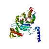 2odnC 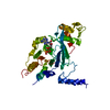 2odwC 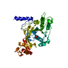 2oe2C 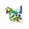 2oesC  2ofoC 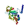 1ubcS S: Starting model for refinement C: citing same article ( |
|---|---|
| Similar structure data |

-Links
 PDBj
PDBj
- Assembly
| Deposited unit | 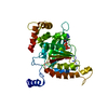
| ||||||||
|---|---|---|---|---|---|---|---|---|---|
| 1 |
| ||||||||
| Unit cell |
|
-Components
| #1: Protein | Mass: 37344.488 Da / Num. of mol.: 1 Source method: isolated from a genetically manipulated source Source: (gene. exp.) Mycobacterium smegmatis (bacteria) / Plasmid: PTHIOA / Production host: References: UniProt: Q59560, UniProt: Q7X416*PLUS, EC: 3.4.99.37 |
|---|---|
| #2: Chemical | ChemComp-ADP /   Mass: 427.201 Da / Num. of mol.: 1 / Source method: obtained synthetically / Formula: C10H15N5O10P2 / Comment: ADP, energy-carrying molecule*YM Mass: 427.201 Da / Num. of mol.: 1 / Source method: obtained synthetically / Formula: C10H15N5O10P2 / Comment: ADP, energy-carrying molecule*YM |
| #3: Water | ChemComp-HOH /  Mass: 18.015 Da / Num. of mol.: 55 / Source method: isolated from a natural source / Formula: H2O Mass: 18.015 Da / Num. of mol.: 55 / Source method: isolated from a natural source / Formula: H2O |
-Experimental details
-Experiment
| Experiment | Method: X-RAY DIFFRACTION / Number of used crystals: 1 |
|---|
- Sample preparation
| Crystal | Density Matthews: 3.3 Å3/Da / Density % sol: 62.75 % |
|---|---|
| Crystal grow | Temperature: 293 K / Method: vapor diffusion, hanging drop / pH: 7.5 Details: Citrate phosphate, sodium citrate, NACL, PEG 4000, pH 7.50, VAPOR DIFFUSION, HANGING DROP, temperature 293K |
-Data collection
| Diffraction | Mean temperature: 293 K |
|---|---|
| Diffraction source | Source: ROTATING ANODE / Type: RIGAKU RU200 / Wavelength: 1.5418 / Wavelength: 1.5418 Å |
| Detector | Type: MARRESEARCH / Detector: IMAGE PLATE / Date: Jan 8, 2003 |
| Radiation | Protocol: SINGLE WAVELENGTH / Monochromatic (M) / Laue (L): M / Scattering type: x-ray |
| Radiation wavelength | Wavelength: 1.5418 Å / Relative weight: 1 |
| Reflection | Resolution: 3.1→21.6 Å / Num. obs: 8712 / % possible obs: 97.3 % / Observed criterion σ(I): 0 / Redundancy: 6.5 % / Biso Wilson estimate: 77.6 Å2 / Rmerge(I) obs: 0.099 / Net I/σ(I): 6.3 |
| Reflection shell | Resolution: 3.1→3.21 Å / Redundancy: 6.5 % / Rmerge(I) obs: 0.539 / Mean I/σ(I) obs: 1.5 / % possible all: 98.6 |
- Processing
| Software |
| ||||||||||||||||||||
|---|---|---|---|---|---|---|---|---|---|---|---|---|---|---|---|---|---|---|---|---|---|
| Refinement | Method to determine structure: MOLECULAR REPLACEMENT Starting model: 1UBC Resolution: 3.1→21.59 Å / Rfactor Rfree error: 0.009 / Data cutoff high absF: 310582.4 / Data cutoff low absF: 0 / Isotropic thermal model: GROUP / Cross valid method: THROUGHOUT / σ(F): 0 / Details: OTHER REFINEMENT REMARKS: NULL
| ||||||||||||||||||||
| Solvent computation | Solvent model: FLAT MODEL / Bsol: 84.6269 Å2 / ksol: 0.271418 e/Å3 | ||||||||||||||||||||
| Displacement parameters | Biso mean: 51 Å2
| ||||||||||||||||||||
| Refine analyze |
| ||||||||||||||||||||
| Refinement step | Cycle: LAST / Resolution: 3.1→21.59 Å
| ||||||||||||||||||||
| Refine LS restraints |
| ||||||||||||||||||||
| LS refinement shell | Resolution: 3.1→3.29 Å / Rfactor Rfree error: 0.033 / Total num. of bins used: 6
| ||||||||||||||||||||
| Xplor file |
|
