| 登録情報 | データベース: PDB / ID: 2odv
|
|---|
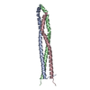
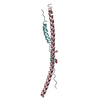
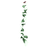
| タイトル | Crystal structure of a fragment of the plakin domain of plectin, Cys to Ala mutant. |
|---|
 要素 要素 | Plectin 1 |
|---|
 キーワード キーワード | STRUCTURAL PROTEIN / PLAKIN DOMAIN / SPECTRIN REPEAT / CYTOSKELETON / HEMIDESMOSOMES / EPIDERMOLYSIS BULLOSA |
|---|
| 機能・相同性 |  機能・相同性情報 機能・相同性情報
protein-containing complex organization / actomyosin contractile ring assembly actin filament organization / tight junction organization / Type I hemidesmosome assembly / skeletal myofibril assembly / hemidesmosome assembly / hemidesmosome / leukocyte migration involved in immune response / intermediate filament organization / intermediate filament cytoskeleton organization ...protein-containing complex organization / actomyosin contractile ring assembly actin filament organization / tight junction organization / Type I hemidesmosome assembly / skeletal myofibril assembly / hemidesmosome assembly / hemidesmosome / leukocyte migration involved in immune response / intermediate filament organization / intermediate filament cytoskeleton organization / dystroglycan binding / fibroblast migration / cellular response to hydrostatic pressure / adherens junction organization / regulation of vascular permeability / costamere / T cell chemotaxis / cellular response to fluid shear stress / intermediate filament cytoskeleton / peripheral nervous system myelin maintenance / myoblast differentiation / cardiac muscle cell development / podosome / structural constituent of muscle / ankyrin binding / Assembly of collagen fibrils and other multimeric structures / sarcomere organization / response to food / nucleus organization / keratinocyte development / transmission of nerve impulse / brush border / sarcoplasm / Caspase-mediated cleavage of cytoskeletal proteins / establishment of skin barrier / skeletal muscle fiber development / respiratory electron transport chain / mitochondrion organization / wound healing / sarcolemma / cell morphogenesis / structural constituent of cytoskeleton / multicellular organism growth / cellular response to mechanical stimulus / Z disc / actin filament binding / intracellular protein localization / myelin sheath / gene expression / mitochondrial outer membrane / cadherin binding / axon / focal adhesion / dendrite / perinuclear region of cytoplasm / RNA binding / extracellular exosome / identical protein binding / plasma membrane / cytosol類似検索 - 分子機能 Spectrin repeat / : / Spectrin-like repeat / Desmoplakin, spectrin-like domain / Spectrin like domain / Plectin repeat / Plectin repeat / Plakin repeat superfamily / Desmoplakin, SH3 domain / SH3 domain ...Spectrin repeat / : / Spectrin-like repeat / Desmoplakin, spectrin-like domain / Spectrin like domain / Plectin repeat / Plectin repeat / Plakin repeat superfamily / Desmoplakin, SH3 domain / SH3 domain / Spectrin-like repeat / Plectin repeat / Plakin / Methane Monooxygenase Hydroxylase; Chain G, domain 1 - #60 / Spectrin/alpha-actinin / Spectrin repeats / Actinin-type actin-binding domain signature 1. / Actinin-type actin-binding domain signature 2. / Actinin-type actin-binding domain, conserved site / Calponin homology domain / Calponin homology (CH) domain / Calponin homology domain / CH domain superfamily / Calponin homology (CH) domain profile. / Plectin/S10, N-terminal / Plectin/S10 domain / Methane Monooxygenase Hydroxylase; Chain G, domain 1 / Src homology 3 (SH3) domain profile. / SH3 domain / Winged helix-like DNA-binding domain superfamily / Up-down Bundle / Mainly Alpha類似検索 - ドメイン・相同性 |
|---|
| 生物種 |  Homo sapiens (ヒト) Homo sapiens (ヒト) |
|---|
| 手法 |  X線回折 / シンクロトロン / 多波長異常分散 / 解像度: 2.05 Å X線回折 / シンクロトロン / 多波長異常分散 / 解像度: 2.05 Å |
|---|
 データ登録者 データ登録者 | de Pereda, J.M. |
|---|
 引用 引用 | ジャーナル: J.Mol.Biol. / 年: 2007
タイトル: The structure of a tandem pair of spectrin repeats of plectin reveals a modular organization of the plakin domain.
著者: Sonnenberg, A. / Rojas, A.M. / de Pereda, J.M. |
|---|
| 履歴 | | 登録 | 2006年12月27日 | 登録サイト: RCSB / 処理サイト: RCSB |
|---|
| 改定 1.0 | 2007年3月13日 | Provider: repository / タイプ: Initial release |
|---|
| 改定 1.1 | 2008年5月1日 | Group: Version format compliance |
|---|
| 改定 1.2 | 2011年7月13日 | Group: Advisory / Version format compliance |
|---|
| 改定 1.3 | 2021年10月20日 | Group: Database references / Derived calculations / カテゴリ: database_2 / struct_ref_seq_dif / struct_site
Item: _database_2.pdbx_DOI / _database_2.pdbx_database_accession ..._database_2.pdbx_DOI / _database_2.pdbx_database_accession / _struct_ref_seq_dif.details / _struct_site.pdbx_auth_asym_id / _struct_site.pdbx_auth_comp_id / _struct_site.pdbx_auth_seq_id |
|---|
| 改定 1.4 | 2023年12月27日 | Group: Data collection / カテゴリ: chem_comp_atom / chem_comp_bond |
|---|
|
|---|
 ムービー
ムービー コントローラー


 データを開く
 基本情報
基本情報 構造の表示
構造の表示 ダウンロードとリンク
 その他のダウンロード
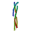


 PDBj
PDBj


 集合体
集合体


 分子量: 76.094 Da / 分子数: 1 / 由来タイプ: 合成 / 式: C3H8O2
 分子量: 18.015 Da / 分子数: 52 / 由来タイプ: 天然 / 式: H2O
 試料調製
試料調製 / ビームライン: BM14 / 波長: 0.9785, 0.9185
 解析
解析