| ソフトウェア | | 名称 | バージョン | 分類 |
|---|
| SHELX | | モデル構築 | | REFMAC | 5.2.0019| 精密化 | | MxCuBE | | データ収集 | | XDS | | データ削減 | | XSCALE | | データスケーリング | | SHELX | | 位相決定 | |
|
|---|
| 精密化 | 構造決定の手法:  多波長異常分散 / 解像度: 2.3→46.47 Å / Cor.coef. Fo:Fc: 0.931 / Cor.coef. Fo:Fc free: 0.899 / SU B: 17.497 / SU ML: 0.202 / TLS residual ADP flag: LIKELY RESIDUAL / Isotropic thermal model: ISOTROPIC / 交差検証法: THROUGHOUT / ESU R: 0.381 / ESU R Free: 0.262 / 立体化学のターゲット値: MAXIMUM LIKELIHOOD 多波長異常分散 / 解像度: 2.3→46.47 Å / Cor.coef. Fo:Fc: 0.931 / Cor.coef. Fo:Fc free: 0.899 / SU B: 17.497 / SU ML: 0.202 / TLS residual ADP flag: LIKELY RESIDUAL / Isotropic thermal model: ISOTROPIC / 交差検証法: THROUGHOUT / ESU R: 0.381 / ESU R Free: 0.262 / 立体化学のターゲット値: MAXIMUM LIKELIHOOD
| Rfactor | 反射数 | %反射 | Selection details |
|---|
| Rfree | 0.2693 | 533 | 4.7 % | RANDOM |
|---|
| Rwork | 0.21616 | - | - | - |
|---|
| obs | 0.21873 | 10704 | 99.94 % | - |
|---|
| all | - | 11237 | - | - |
|---|
|
|---|
| 溶媒の処理 | イオンプローブ半径: 0.8 Å / 減衰半径: 0.8 Å / VDWプローブ半径: 1.4 Å / 溶媒モデル: BABINET MODEL WITH MASK |
|---|
| 原子変位パラメータ | Biso mean: 40.371 Å2
| Baniso -1 | Baniso -2 | Baniso -3 |
|---|
| 1- | -1.01 Å2 | 0 Å2 | 0 Å2 |
|---|
| 2- | - | -1.33 Å2 | 0 Å2 |
|---|
| 3- | - | - | 2.35 Å2 |
|---|
|
|---|
| 精密化ステップ | サイクル: LAST / 解像度: 2.3→46.47 Å
| タンパク質 | 核酸 | リガンド | 溶媒 | 全体 |
|---|
| 原子数 | 1795 | 0 | 0 | 16 | 1811 |
|---|
|
|---|
| 拘束条件 | | Refine-ID | タイプ | Dev ideal | Dev ideal target | 数 |
|---|
| X-RAY DIFFRACTION | r_bond_refined_d| 0.015 | 0.022 | 1879 | | X-RAY DIFFRACTION | r_bond_other_d | | | | X-RAY DIFFRACTION | r_angle_refined_deg| 1.564 | 1.972 | 2532 | | X-RAY DIFFRACTION | r_angle_other_deg | | | | X-RAY DIFFRACTION | r_dihedral_angle_1_deg| 5.44 | 5 | 225 | | X-RAY DIFFRACTION | r_dihedral_angle_2_deg| 33.551 | 23.564 | 101 | | X-RAY DIFFRACTION | r_dihedral_angle_3_deg| 20.161 | 15 | 369 | | X-RAY DIFFRACTION | r_dihedral_angle_4_deg| 14.932 | 15 | 21 | | X-RAY DIFFRACTION | r_chiral_restr| 0.102 | 0.2 | 268 | | X-RAY DIFFRACTION | r_gen_planes_refined| 0.006 | 0.02 | 1437 | | X-RAY DIFFRACTION | r_gen_planes_other | | | | X-RAY DIFFRACTION | r_nbd_refined| 0.224 | 0.2 | 901 | | X-RAY DIFFRACTION | r_nbd_other | | | | X-RAY DIFFRACTION | r_nbtor_refined| 0.3 | 0.2 | 1301 | | X-RAY DIFFRACTION | r_nbtor_other | | | | X-RAY DIFFRACTION | r_xyhbond_nbd_refined| 0.145 | 0.2 | 55 | | X-RAY DIFFRACTION | r_xyhbond_nbd_other | | | | X-RAY DIFFRACTION | r_metal_ion_refined | | | | X-RAY DIFFRACTION | r_metal_ion_other | | | | X-RAY DIFFRACTION | r_symmetry_vdw_refined| 0.276 | 0.2 | 60 | | X-RAY DIFFRACTION | r_symmetry_vdw_other | | | | X-RAY DIFFRACTION | r_symmetry_hbond_refined| 0.253 | 0.2 | 9 | | X-RAY DIFFRACTION | r_symmetry_hbond_other | | | | X-RAY DIFFRACTION | r_symmetry_metal_ion_refined | | | | X-RAY DIFFRACTION | r_symmetry_metal_ion_other | | | | X-RAY DIFFRACTION | r_mcbond_it| 0.874 | 1.5 | 1150 | | X-RAY DIFFRACTION | r_mcbond_other | | | | X-RAY DIFFRACTION | r_mcangle_it| 1.362 | 2 | 1783 | | X-RAY DIFFRACTION | r_scbond_it| 2.374 | 3 | 825 | | X-RAY DIFFRACTION | r_scangle_it| 3.677 | 4.5 | 749 | | X-RAY DIFFRACTION | r_rigid_bond_restr | | | | X-RAY DIFFRACTION | r_sphericity_free | | | | X-RAY DIFFRACTION | r_sphericity_bonded | | | | | | | | | | | | | | | | | | | | | | | | | | | | | | | | | | | |
|
|---|
| LS精密化 シェル | 解像度: 2.3→2.36 Å / Total num. of bins used: 20
| Rfactor | 反射数 | %反射 |
|---|
| Rfree | 0.356 | 42 | - |
|---|
| Rwork | 0.208 | 769 | - |
|---|
| obs | - | - | 100 % |
|---|
|
|---|
| 精密化 TLS | 手法: refined / Refine-ID: X-RAY DIFFRACTION | ID | L11 (°2) | L12 (°2) | L13 (°2) | L22 (°2) | L23 (°2) | L33 (°2) | S11 (Å °) | S12 (Å °) | S13 (Å °) | S21 (Å °) | S22 (Å °) | S23 (Å °) | S31 (Å °) | S32 (Å °) | S33 (Å °) | T11 (Å2) | T12 (Å2) | T13 (Å2) | T22 (Å2) | T23 (Å2) | T33 (Å2) | Origin x (Å) | Origin y (Å) | Origin z (Å) |
|---|
| 1 | 5.5023 | -4.7606 | 10.9328 | 5.9224 | -9.6767 | 22.7471 | -0.3641 | 0.5668 | 0.5925 | -0.1584 | -0.446 | -0.4853 | -1.0718 | 1.0901 | 0.8101 | 0.1551 | -0.0599 | 0.0373 | -0.0475 | 0.045 | -0.0663 | 30.347 | 22.55 | 3.354 | | 2 | 3.2932 | -3.3336 | 5.9498 | 5.3763 | -8.0417 | 14.1236 | 0.344 | -0.0221 | -0.1574 | -0.9098 | 0.1865 | 0.1593 | 0.8146 | -0.5458 | -0.5305 | 0.0511 | -0.0384 | -0.0012 | -0.1108 | 0.0161 | -0.2541 | 24.502 | 17.817 | 0.883 | | 3 | 9.543 | -6.1793 | 2.1134 | 5.5745 | -1.2852 | 3.336 | -0.2859 | -0.7055 | 0.3634 | -0.1112 | 0.256 | -0.3864 | 0.1046 | 0.0058 | 0.0299 | -0.192 | -0.0068 | -0.0195 | -0.1549 | -0.0251 | -0.2339 | 57.616 | -9.209 | 35.156 | | 4 | 21.2422 | -10.9361 | 9.1802 | 9.8051 | -5.032 | 8.9247 | -0.2319 | 0.3806 | 0.5127 | -0.1021 | -0.3747 | -1.325 | 0.0135 | 0.7603 | 0.6066 | -0.1756 | -0.0523 | 0.0352 | 0.0417 | -0.0146 | 0.2652 | 68.577 | -11.844 | 36.146 |
|
|---|
| 精密化 TLSグループ | | ID | Refine-ID | Refine TLS-ID | Auth asym-ID | Label asym-ID | Auth seq-ID | Label seq-ID |
|---|
| 1 | X-RAY DIFFRACTION | 1 | AA| 303 - 346 | 8 - 51 | | 2 | X-RAY DIFFRACTION | 2 | AA| 347 - 417 | 52 - 122 | | 3 | X-RAY DIFFRACTION | 3 | AA| 418 - 499 | 123 - 204 | | 4 | X-RAY DIFFRACTION | 4 | AA| 500 - 520 | 205 - 225 | | | | | | | | |
|
|---|
 ムービー
ムービー コントローラー


 データを開く
 基本情報
基本情報 要素
要素 キーワード
キーワード 機能・相同性情報
 Homo sapiens (ヒト)
 データ登録者
 引用
引用 構造の表示
構造の表示 ダウンロードとリンク
 その他のダウンロード


 PDBj
PDBj


 集合体
集合体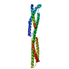

 分子量: 18.015 Da / 分子数: 16 / 由来タイプ: 天然 / 式: H2O
 試料調製
試料調製 / ビームライン: BM14 / 波長: 0.9785, 0.9185
 解析
解析