SEQUENCE AUTHORS STATE THAT THE DIFFERENCE BETWEEN THEIR CLONED SEQUENCE AND THE DATABASE SEQUENCE ... SEQUENCE AUTHORS STATE THAT THE DIFFERENCE BETWEEN THEIR CLONED SEQUENCE AND THE DATABASE SEQUENCE AT POSITION 457 REPRESENTS VARIATION IN THE PHAGE SEQUENCE AND NOT A MUTATION INCORPORATED BY AMPLIFICATION OF THE GENE.
Resolution: 2.7→30 Å / FOM work R set: 0.821 / Isotropic thermal model: Restrained / Cross valid method: THROUGHOUT / σ(F): 0 / Stereochemistry target values: Engh & Huber Details: Atoms at 0.00 occupancy have little or no electron density and were modelled stereochemically. Residues 287-295 are missing due to insufficient electron density
Rfactor
Num. reflection
% reflection
Selection details
Rfree
0.25
1292
5 %
Random
Rwork
0.217
-
-
-
obs
-
25425
97.5 %
-
Solvent computation
Bsol: 50.771 Å2
Displacement parameters
Biso mean: 69.1 Å2
Baniso -1
Baniso -2
Baniso -3
1-
-14.783 Å2
0 Å2
0 Å2
2-
-
30.546 Å2
0 Å2
3-
-
-
-15.762 Å2
Refine analyze
Free
Obs
Luzzati coordinate error
0.38 Å
0.32 Å
Luzzati d res low
-
5 Å
Luzzati sigma a
0.48 Å
0.44 Å
Refinement step
Cycle: LAST / Resolution: 2.7→30 Å
Protein
Nucleic acid
Ligand
Solvent
Total
Num. atoms
4017
0
0
106
4123
Refine LS restraints
Refine-ID
Type
Dev ideal
Dev ideal target
X-RAY DIFFRACTION
c_bond_d
0.007
X-RAY DIFFRACTION
c_angle_deg
1.261
X-RAY DIFFRACTION
c_mcbond_it
1.344
1.5
X-RAY DIFFRACTION
c_scbond_it
2.018
2
X-RAY DIFFRACTION
c_mcangle_it
2.331
2
X-RAY DIFFRACTION
c_scangle_it
3.126
2.5
X-RAY DIFFRACTION
c_dihedral_angle_d
22.8
X-RAY DIFFRACTION
c_improper_angle_d
0.68
LS refinement shell
Refine-ID: X-RAY DIFFRACTION / Total num. of bins used: 25
Resolution (Å)
Rfactor Rfree
Num. reflection Rfree
Rfactor Rwork
Num. reflection Rwork
Num. reflection obs
2.7-2.74
0.393
38
0.342
756
794
2.74-2.78
0.325
57
0.319
826
883
2.78-2.82
0.362
56
0.326
862
918
2.82-2.86
0.294
51
0.308
895
946
2.86-2.91
0.291
44
0.281
914
958
2.91-2.96
0.315
50
0.267
977
1027
2.96-3.01
0.299
40
0.266
969
1009
3.01-3.07
0.304
60
0.257
991
1051
3.07-3.13
0.248
54
0.249
983
1037
3.13-3.2
0.373
56
0.252
975
1031
3.2-3.28
0.321
62
0.233
974
1036
3.28-3.36
0.254
63
0.233
957
1020
3.36-3.45
0.224
59
0.207
975
1034
3.45-3.55
0.228
68
0.197
970
1038
3.55-3.66
0.216
41
0.184
986
1027
3.66-3.79
0.202
53
0.185
1002
1055
3.79-3.95
0.23
56
0.188
981
1037
3.95-4.12
0.275
44
0.193
999
1043
4.12-4.34
0.201
62
0.184
976
1038
4.34-4.61
0.21
58
0.169
1000
1058
4.61-4.97
0.194
43
0.179
1006
1049
4.97-5.46
0.259
46
0.199
1014
1060
5.46-6.25
0.361
49
0.266
1020
1069
6.25-7.85
0.24
41
0.246
1040
1081
7.85-30
0.221
41
0.225
1085
1126
Xplor file
Refine-ID
Serial no
Param file
Topol file
X-RAY DIFFRACTION
1
CNS_TOPPAR:protein_rep.param
CNS_TOPPAR:protein.top
X-RAY DIFFRACTION
2
CNS_TOPPAR:dna-rna_rep.param
CNS_TOPPAR:dna-rna.top
X-RAY DIFFRACTION
3
CNS_TOPPAR:water_rep.param
CNS_TOPPAR:water.top
X-RAY DIFFRACTION
4
CNS_TOPPAR:ion.param
CNS_TOPPAR:ion.top
+
About Yorodumi
-
News
-
Feb 9, 2022. New format data for meta-information of EMDB entries
New format data for meta-information of EMDB entries
Version 3 of the EMDB header file is now the official format.
The previous official version 1.9 will be removed from the archive.
In the structure databanks used in Yorodumi, some data are registered as the other names, "COVID-19 virus" and "2019-nCoV". Here are the details of the virus and the list of structure data.
Jan 31, 2019. EMDB accession codes are about to change! (news from PDBe EMDB page)
EMDB accession codes are about to change! (news from PDBe EMDB page)
The allocation of 4 digits for EMDB accession codes will soon come to an end. Whilst these codes will remain in use, new EMDB accession codes will include an additional digit and will expand incrementally as the available range of codes is exhausted. The current 4-digit format prefixed with “EMD-” (i.e. EMD-XXXX) will advance to a 5-digit format (i.e. EMD-XXXXX), and so on. It is currently estimated that the 4-digit codes will be depleted around Spring 2019, at which point the 5-digit format will come into force.
The EM Navigator/Yorodumi systems omit the EMD- prefix.
Related info.:Q: What is EMD? / ID/Accession-code notation in Yorodumi/EM Navigator
Yorodumi is a browser for structure data from EMDB, PDB, SASBDB, etc.
This page is also the successor to EM Navigator detail page, and also detail information page/front-end page for Omokage search.
The word "yorodu" (or yorozu) is an old Japanese word meaning "ten thousand". "mi" (miru) is to see.
Related info.:EMDB / PDB / SASBDB / Comparison of 3 databanks / Yorodumi Search / Aug 31, 2016. New EM Navigator & Yorodumi / Yorodumi Papers / Jmol/JSmol / Function and homology information / Changes in new EM Navigator and Yorodumi
 Movie
Movie Controller


 Open data
 Basic information
 Components
 Keywords
 Function and homology information
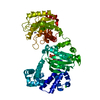
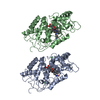
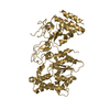
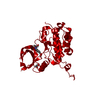
 Enterobacteria phage T4 (virus)
 X-RAY DIFFRACTION /
  Authors
 Citation
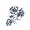
 Structure visualization
 Downloads & links
 Other downloads


 PDBj
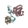
PDBj Assembly
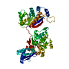

 Mass: 18.015 Da / Num. of mol.: 106 / Source method: isolated from a natural source / Formula: H2O
 Sample preparation
 / Beamline: 22-ID / Wavelength: 1.05 Å
 Processing