SEQUENCE THE CONSTRUCT WAS EXPRESSED WITH A PURIFICATION TAG MGSDKIHHHHHHENLYFQG. THE TAG WAS ...SEQUENCE THE CONSTRUCT WAS EXPRESSED WITH A PURIFICATION TAG MGSDKIHHHHHHENLYFQG. THE TAG WAS REMOVED WITH TEV PROTEASE LEAVING ONLY A GLYCINE (0) FOLLOWED BY THE TARGET SEQUENCE.
Type: MARMOSAIC 325 mm CCD / Detector: CCD / Date: Nov 19, 2006 / Details: Flat mirror (vertical focusing)
Radiation
Monochromator: Single crystal Si(111) bent monochromator (horizontal focusing) Protocol: MAD / Monochromatic (M) / Laue (L): M / Scattering type: x-ray
Radiation wavelength
ID
Wavelength (Å)
Relative weight
1
0.91837
1
2
0.97978
1
Reflection
Resolution: 1.5→28.262 Å / Num. obs: 29534 / % possible obs: 94.8 % / Redundancy: 3.71 % / Biso Wilson estimate: 12.94 Å2 / Rmerge(I) obs: 0.046 / Net I/σ(I): 11.11
Reflection shell
Diffraction-ID: 1
Resolution (Å)
Highest resolution (Å)
Rmerge(I) obs
Mean I/σ(I) obs
Num. measured obs
Num. unique all
% possible all
1.5-1.55
0.271
2.88
8560
4527
88.8
1.55-1.62
0.208
3.9
11711
5856
88.6
1.62-1.69
0.167
4.8
9909
4970
90.7
1.69-1.78
0.127
6
10749
5387
91.3
1.78-1.89
0.099
7.7
10661
5343
92.2
1.89-2.04
0.069
10.5
11124
5594
93.8
2.04-2.24
0.051
13.5
10756
5404
95
2.24-2.56
0.038
16.5
11028
5546
95.6
2.56
0.034
19.9
11221
5690
96.7
-
Phasing
Phasing
Method: MAD
-
Processing
Software
Name
Version
Classification
NB
MolProbity
3beta29
modelbuilding
SHELX
phasing
REFMAC
5.2.0005
refinement
XSCALE
datascaling
PDB_EXTRACT
2
dataextraction
XDS
datareduction
SHELXD
phasing
autoSHARP
phasing
Refinement
Method to determine structure: MAD / Resolution: 1.5→28.262 Å / Cor.coef. Fo:Fc: 0.975 / Cor.coef. Fo:Fc free: 0.965 / SU B: 1.152 / SU ML: 0.044 / Cross valid method: THROUGHOUT / σ(F): 0 / ESU R: 0.065 / ESU R Free: 0.067 Stereochemistry target values: MAXIMUM LIKELIHOOD WITH PHASES Details: 1. HYDROGENS HAVE BEEN ADDED IN THE RIDING POSITIONS. 2. A MET-INHIBITION PROTOCOL WAS USED FOR SELENOMETHIONINE INCORPORATION DURING PROTEIN EXPRESSION. THE OCCUPANCY OF THE SE ATOMS IN THE ...Details: 1. HYDROGENS HAVE BEEN ADDED IN THE RIDING POSITIONS. 2. A MET-INHIBITION PROTOCOL WAS USED FOR SELENOMETHIONINE INCORPORATION DURING PROTEIN EXPRESSION. THE OCCUPANCY OF THE SE ATOMS IN THE MSE RESIDUES WAS REDUCED TO 0.75 FOR THE REDUCED SCATTERING POWER DUE TO PARTIAL S-MET INCORPORATION.
Rfactor
Num. reflection
% reflection
Selection details
Rfree
0.167
1499
5.1 %
RANDOM
Rwork
0.138
-
-
-
obs
0.139
29513
99.57 %
-
Solvent computation
Ion probe radii: 0.8 Å / Shrinkage radii: 0.8 Å / VDW probe radii: 1.2 Å / Solvent model: BABINET MODEL WITH MASK
Displacement parameters
Biso mean: 13.085 Å2
Baniso -1
Baniso -2
Baniso -3
1-
0.26 Å2
0 Å2
-0.75 Å2
2-
-
-0.22 Å2
0 Å2
3-
-
-
-0.51 Å2
Refinement step
Cycle: LAST / Resolution: 1.5→28.262 Å
Protein
Nucleic acid
Ligand
Solvent
Total
Num. atoms
1566
0
12
328
1906
Refine LS restraints
Refine-ID
Type
Dev ideal
Dev ideal target
Number
X-RAY DIFFRACTION
r_bond_refined_d
0.014
0.022
1651
X-RAY DIFFRACTION
r_bond_other_d
0.001
0.02
1583
X-RAY DIFFRACTION
r_angle_refined_deg
1.457
1.987
2256
X-RAY DIFFRACTION
r_angle_other_deg
0.826
3
3662
X-RAY DIFFRACTION
r_dihedral_angle_1_deg
5.645
5
219
X-RAY DIFFRACTION
r_dihedral_angle_2_deg
31.741
23.043
69
X-RAY DIFFRACTION
r_dihedral_angle_3_deg
10.84
15
257
X-RAY DIFFRACTION
r_dihedral_angle_4_deg
16.713
15
16
X-RAY DIFFRACTION
r_chiral_restr
0.084
0.2
254
X-RAY DIFFRACTION
r_gen_planes_refined
0.006
0.02
1863
X-RAY DIFFRACTION
r_gen_planes_other
0.001
0.02
326
X-RAY DIFFRACTION
r_nbd_refined
0.207
0.2
316
X-RAY DIFFRACTION
r_nbd_other
0.187
0.2
1638
X-RAY DIFFRACTION
r_nbtor_refined
0.175
0.2
796
X-RAY DIFFRACTION
r_nbtor_other
0.081
0.2
989
X-RAY DIFFRACTION
r_xyhbond_nbd_refined
0.182
0.2
210
X-RAY DIFFRACTION
r_symmetry_vdw_refined
0.105
0.2
8
X-RAY DIFFRACTION
r_symmetry_vdw_other
0.17
0.2
51
X-RAY DIFFRACTION
r_symmetry_hbond_refined
0.189
0.2
36
X-RAY DIFFRACTION
r_mcbond_it
1.72
3
1128
X-RAY DIFFRACTION
r_mcbond_other
0.405
3
430
X-RAY DIFFRACTION
r_mcangle_it
2.337
5
1702
X-RAY DIFFRACTION
r_scbond_it
3.621
8
637
X-RAY DIFFRACTION
r_scangle_it
5.59
11
548
LS refinement shell
Resolution: 1.501→1.54 Å / Total num. of bins used: 20
Rfactor
Num. reflection
% reflection
Rfree
0.238
103
-
Rwork
0.188
2028
-
obs
-
2131
97.48 %
+
About Yorodumi
-
News
-
Feb 9, 2022. New format data for meta-information of EMDB entries
New format data for meta-information of EMDB entries
Version 3 of the EMDB header file is now the official format.
The previous official version 1.9 will be removed from the archive.
In the structure databanks used in Yorodumi, some data are registered as the other names, "COVID-19 virus" and "2019-nCoV". Here are the details of the virus and the list of structure data.
Jan 31, 2019. EMDB accession codes are about to change! (news from PDBe EMDB page)
EMDB accession codes are about to change! (news from PDBe EMDB page)
The allocation of 4 digits for EMDB accession codes will soon come to an end. Whilst these codes will remain in use, new EMDB accession codes will include an additional digit and will expand incrementally as the available range of codes is exhausted. The current 4-digit format prefixed with “EMD-” (i.e. EMD-XXXX) will advance to a 5-digit format (i.e. EMD-XXXXX), and so on. It is currently estimated that the 4-digit codes will be depleted around Spring 2019, at which point the 5-digit format will come into force.
The EM Navigator/Yorodumi systems omit the EMD- prefix.
Related info.:Q: What is EMD? / ID/Accession-code notation in Yorodumi/EM Navigator
Yorodumi is a browser for structure data from EMDB, PDB, SASBDB, etc.
This page is also the successor to EM Navigator detail page, and also detail information page/front-end page for Omokage search.
The word "yorodu" (or yorozu) is an old Japanese word meaning "ten thousand". "mi" (miru) is to see.
Related info.:EMDB / PDB / SASBDB / Comparison of 3 databanks / Yorodumi Search / Aug 31, 2016. New EM Navigator & Yorodumi / Yorodumi Papers / Jmol/JSmol / Function and homology information / Changes in new EM Navigator and Yorodumi
 Movie
Movie Controller

 Yorodumi
 Open data
 Basic information
 Components
 Keywords
 Function and homology information
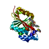
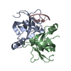
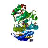
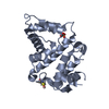
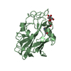
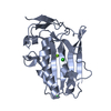
 Mesorhizobium loti (bacteria)
 X-RAY DIFFRACTION /
  Authors
 Citation
 Structure visualization
 Downloads & links
 Other downloads


 PDBj
PDBj Assembly
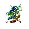

 Mass: 92.094 Da / Num. of mol.: 2 / Source method: obtained synthetically / Formula: C3H8O3
 Mass: 18.015 Da / Num. of mol.: 328 / Source method: isolated from a natural source / Formula: H2O
 Sample preparation
 / Beamline: BL11-1 / Wavelength: 0.91837, 0.97978
 Processing