 Movie
Movie Controller

[English] 日本語
 Yorodumi
- PDB-2o2j: Mycobacterium tuberculosis tryptophan synthase beta chain dimer (... -
+ Open data

- Basic information
| Entry | Database: PDB / ID: 2o2j | ||||||
|---|---|---|---|---|---|---|---|
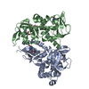
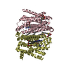
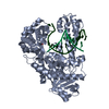
| Title | Mycobacterium tuberculosis tryptophan synthase beta chain dimer (apoform) | ||||||
 Components Components | Tryptophan synthase beta chain | ||||||
 Keywords Keywords | LYASE / Amino-acid biosynthesis / Tryptophan biosynthesis / Structural Genomics / Mycobacterium Tuberculosis Structural Proteomics Project / XMTB | ||||||
| Function / homology |  Function and homology information Function and homology informationtryptophan synthase / tryptophan synthase activity / L-tryptophan biosynthetic process / cytoplasm Similarity search - Function | ||||||
| Biological species |   Mycobacterium tuberculosis (bacteria) Mycobacterium tuberculosis (bacteria) | ||||||
| Method |  X-RAY DIFFRACTION / SYNCHROTRON / MOLECULAR REPLACEMENT / Resolution: 2.56 Å X-RAY DIFFRACTION / SYNCHROTRON / MOLECULAR REPLACEMENT / Resolution: 2.56 Å | ||||||
 Authors Authors | Burenkov, G.P. / Kachalova, G.S. / Bartunik, H.D. / Mycobacterium Tuberculosis Structural Proteomics Project (XMTB) | ||||||
 Citation Citation | Journal: To be Published Title: Structure Tryptophan SYNTHASE beta chain dimer from MYCOBACTERIUM Tuberculosis Authors: Kachalova, G.S. / Burenkov, G.P. / I Strizhov, N. / I Svergun, D. / Bartunik, H.D. | ||||||
| History |
|
- Structure visualization
| Structure viewer | Molecule: MolmilJmol/JSmol |
|---|
- Downloads & links
-Download
| PDBx/mmCIF format | 2o2j.cif.gz | 132.8 KB | Display | PDBx/mmCIF format |
|---|---|---|---|---|
| PDB format | pdb2o2j.ent.gz | 102.5 KB | Display | PDB format |
| PDBx/mmJSON format | 2o2j.json.gz | Tree view | PDBx/mmJSON format | |
| Others |  Other downloads Other downloads |
-Validation report
| Arichive directory | https://data.pdbj.org/pub/pdb/validation_reports/o2/2o2jftp://data.pdbj.org/pub/pdb/validation_reports/o2/2o2j | HTTPS FTP |
|---|
-Related structure data
| Related structure data | 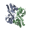 2o2eS S: Starting model for refinement |
|---|---|
| Similar structure data | |
| Other databases |

-Links
 PDBj
PDBj
- Assembly
| Deposited unit | 
| ||||||||
|---|---|---|---|---|---|---|---|---|---|
| 1 |
| ||||||||
| Unit cell |
|
-Components
| #1: Protein | Mass: 44694.164 Da / Num. of mol.: 2 Source method: isolated from a genetically manipulated source Source: (gene. exp.) Mycobacterium tuberculosis (bacteria) / Gene: trpB / Plasmid: p6d, pET24b / Production host: References: UniProt: P66984, UniProt: P9WFX9*PLUS, tryptophan synthase #2: Water | ChemComp-HOH / |  Mass: 18.015 Da / Num. of mol.: 20 / Source method: isolated from a natural source / Formula: H2O Mass: 18.015 Da / Num. of mol.: 20 / Source method: isolated from a natural source / Formula: H2O |
|---|
-Experimental details
-Experiment
| Experiment | Method: X-RAY DIFFRACTION / Number of used crystals: 1 |
|---|
- Sample preparation
| Crystal | Density Matthews: 2.15 Å3/Da / Density % sol: 42.67 % |
|---|---|
| Crystal grow | Temperature: 293 K / Method: vapor diffusion, sitting drop / pH: 7.5 Details: 0.1 M Hepes, 2.0 M Ammonium Formate, pH 7.5, VAPOR DIFFUSION, SITTING DROP, temperature 293K |
-Data collection
| Diffraction | Mean temperature: 100 K |
|---|---|
| Diffraction source | Source: SYNCHROTRON / Site: MPG/DESY, HAMBURG  / Beamline: BW6 / Wavelength: 1.05 Å / Beamline: BW6 / Wavelength: 1.05 Å |
| Detector | Type: MAR CCD 165 mm / Detector: CCD / Date: Feb 26, 2004 / Details: Au-coated planar; toroidal mirrors |
| Radiation | Monochromator: Si111 / Protocol: SINGLE WAVELENGTH / Monochromatic (M) / Laue (L): M / Scattering type: x-ray |
| Radiation wavelength | Wavelength: 1.05 Å / Relative weight: 1 |
| Reflection | Resolution: 2.56→30 Å / Num. all: 23336 / Num. obs: 22608 / % possible obs: 96.9 % / Observed criterion σ(I): 2 / Redundancy: 2 % / Rmerge(I) obs: 0.047 / Net I/σ(I): 13.5 |
| Reflection shell | Resolution: 2.56→2.63 Å / Redundancy: 1.8 % / Rmerge(I) obs: 0.367 / Mean I/σ(I) obs: 2 / Num. unique all: 1622 / % possible all: 93.4 |
- Processing
| Software |
| ||||||||||||||||||||||||||||||||||||||||||||||||||||||||||||||||||||||||||||||||||||||||||
|---|---|---|---|---|---|---|---|---|---|---|---|---|---|---|---|---|---|---|---|---|---|---|---|---|---|---|---|---|---|---|---|---|---|---|---|---|---|---|---|---|---|---|---|---|---|---|---|---|---|---|---|---|---|---|---|---|---|---|---|---|---|---|---|---|---|---|---|---|---|---|---|---|---|---|---|---|---|---|---|---|---|---|---|---|---|---|---|---|---|---|---|
| Refinement | Method to determine structure: MOLECULAR REPLACEMENT Starting model: PDB ENTRY 2O2E Resolution: 2.56→10 Å / Cor.coef. Fo:Fc: 0.941 / Cor.coef. Fo:Fc free: 0.876 / SU B: 12.016 / SU ML: 0.266 / Isotropic thermal model: isotropic / Cross valid method: THROUGHOUT / ESU R: 0.638 / ESU R Free: 0.367 / Stereochemistry target values: MAXIMUM LIKELIHOOD
| ||||||||||||||||||||||||||||||||||||||||||||||||||||||||||||||||||||||||||||||||||||||||||
| Solvent computation | Ion probe radii: 0.8 Å / Shrinkage radii: 0.8 Å / VDW probe radii: 1.2 Å / Solvent model: BABINET MODEL WITH MASK | ||||||||||||||||||||||||||||||||||||||||||||||||||||||||||||||||||||||||||||||||||||||||||
| Displacement parameters | Biso mean: 45.799 Å2
| ||||||||||||||||||||||||||||||||||||||||||||||||||||||||||||||||||||||||||||||||||||||||||
| Refinement step | Cycle: LAST / Resolution: 2.56→10 Å
| ||||||||||||||||||||||||||||||||||||||||||||||||||||||||||||||||||||||||||||||||||||||||||
| Refine LS restraints |
| ||||||||||||||||||||||||||||||||||||||||||||||||||||||||||||||||||||||||||||||||||||||||||
| LS refinement shell | Resolution: 2.56→2.626 Å / Total num. of bins used: 20
|
