 Movie
Movie Controller

[English] 日本語
 Yorodumi
- PDB-2nzf: Structure of beta-lactamase II from Bacillus cereus. R121H, C221S... -
+ Open data

- Basic information
| Entry | Database: PDB / ID: 2nzf | ||||||
|---|---|---|---|---|---|---|---|
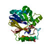
| Title | Structure of beta-lactamase II from Bacillus cereus. R121H, C221S double mutant. Space group C2. | ||||||
 Components Components | Beta-lactamase II | ||||||
 Keywords Keywords | HYDROLASE / Beta-Lactamase II / Bacillus cereus | ||||||
| Function / homology |  Function and homology information Function and homology informationantibiotic catabolic process / beta-lactamase activity / beta-lactamase / periplasmic space / response to antibiotic / zinc ion binding Similarity search - Function | ||||||
| Biological species |  | ||||||
| Method |  X-RAY DIFFRACTION / SYNCHROTRON / MOLECULAR REPLACEMENT / Resolution: 2.28 Å X-RAY DIFFRACTION / SYNCHROTRON / MOLECULAR REPLACEMENT / Resolution: 2.28 Å | ||||||
 Authors Authors | Medrano Martin, F.J. / Vila, A.J. / Gonzalez, J.M. | ||||||
 Citation Citation | Journal: J.Mol.Biol. / Year: 2007 Title: The Zn2 position in metallo-beta-lactamases is critical for activity: a study on chimeric metal sites on a conserved protein scaffold. Authors: Gonzalez, J.M. / Medrano Martin, F.J. / Costello, A.L. / Tierney, D.L. / Vila, A.J. | ||||||
| History |
|
- Structure visualization
| Structure viewer | Molecule: MolmilJmol/JSmol |
|---|
- Downloads & links
-Download
| PDBx/mmCIF format | 2nzf.cif.gz | 58.7 KB | Display | PDBx/mmCIF format |
|---|---|---|---|---|
| PDB format | pdb2nzf.ent.gz | 41.2 KB | Display | PDB format |
| PDBx/mmJSON format | 2nzf.json.gz | Tree view | PDBx/mmJSON format | |
| Others |  Other downloads Other downloads |
-Validation report
| Summary document | 2nzf_validation.pdf.gz | 427.2 KB | Display | wwPDB validaton report |
|---|---|---|---|---|
| Full document | 2nzf_full_validation.pdf.gz | 429.6 KB | Display | |
| Data in XML | 2nzf_validation.xml.gz | 11.5 KB | Display | |
| Data in CIF | 2nzf_validation.cif.gz | 15.9 KB | Display | |
| Arichive directory | https://data.pdbj.org/pub/pdb/validation_reports/nz/2nzfftp://data.pdbj.org/pub/pdb/validation_reports/nz/2nzf | HTTPS FTP |
-Related structure data
| Related structure data |  2nxaC  2nypC  2nzeC  1bc2S S: Starting model for refinement C: citing same article ( |
|---|---|
| Similar structure data |


-Links
 PDBj
PDBj

- Assembly
| Deposited unit | 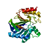
| ||||||||
|---|---|---|---|---|---|---|---|---|---|
| 1 |
| ||||||||
| Unit cell |
|
-Components
| #1: Protein | Mass: 24258.611 Da / Num. of mol.: 1 Source method: isolated from a genetically manipulated source Source: (gene. exp.) |
|---|---|
| #2: Chemical | ChemComp-ZN /   Mass: 65.409 Da / Num. of mol.: 1 / Source method: obtained synthetically / Formula: Zn Mass: 65.409 Da / Num. of mol.: 1 / Source method: obtained synthetically / Formula: Zn |
| #3: Water | ChemComp-HOH /  Mass: 18.015 Da / Num. of mol.: 130 / Source method: isolated from a natural source / Formula: H2O Mass: 18.015 Da / Num. of mol.: 130 / Source method: isolated from a natural source / Formula: H2O |
-Experimental details
-Experiment
| Experiment | Method: X-RAY DIFFRACTION / Number of used crystals: 1 |
|---|
- Sample preparation
| Crystal | Density Matthews: 2.27 Å3/Da / Density % sol: 45.84 % |
|---|---|
| Crystal grow | Temperature: 295 K / Method: vapor diffusion, hanging drop / pH: 5.4 Details: 0.1 M sodium cacodylate, 60 mM sodium tartrate, 18% PEG 3350, pH 5.4, VAPOR DIFFUSION, HANGING DROP, temperature 295K |
-Data collection
| Diffraction | Mean temperature: 100 K |
|---|---|
| Diffraction source | Source: SYNCHROTRON / Site: LNLS  / Beamline: D03B-MX1 / Beamline: D03B-MX1 |
| Detector | Type: MAR CCD 165 mm / Detector: CCD / Date: Feb 15, 2006 |
| Radiation | Protocol: SINGLE WAVELENGTH / Monochromatic (M) / Laue (L): M / Scattering type: x-ray |
| Radiation wavelength | Relative weight: 1 |
| Reflection | Resolution: 2.28→50 Å / Num. obs: 9968 / % possible obs: 98.3 % / Redundancy: 6.4 % / Rsym value: 0.059 / Net I/σ(I): 14.8 |
| Reflection shell | Resolution: 2.28→2.35 Å / Redundancy: 2.8 % / Mean I/σ(I) obs: 9.7 / Rsym value: 0.17 / % possible all: 83 |
- Processing
| Software |
| |||||||||||||||||||||||||||||||||||||||||||||||||||||||||||||||||||||||||||||||||||||||||||||||
|---|---|---|---|---|---|---|---|---|---|---|---|---|---|---|---|---|---|---|---|---|---|---|---|---|---|---|---|---|---|---|---|---|---|---|---|---|---|---|---|---|---|---|---|---|---|---|---|---|---|---|---|---|---|---|---|---|---|---|---|---|---|---|---|---|---|---|---|---|---|---|---|---|---|---|---|---|---|---|---|---|---|---|---|---|---|---|---|---|---|---|---|---|---|---|---|---|
| Refinement | Method to determine structure: MOLECULAR REPLACEMENT Starting model: 1bc2 Resolution: 2.28→68.52 Å / Cor.coef. Fo:Fc: 0.954 / Cor.coef. Fo:Fc free: 0.908 / SU B: 6.257 / SU ML: 0.156 / Cross valid method: THROUGHOUT / σ(F): 0 / ESU R: 0.335 / ESU R Free: 0.241 / Stereochemistry target values: MAXIMUM LIKELIHOOD / Details: HYDROGENS HAVE BEEN ADDED IN THE RIDING POSITIONS
| |||||||||||||||||||||||||||||||||||||||||||||||||||||||||||||||||||||||||||||||||||||||||||||||
| Solvent computation | Ion probe radii: 0.8 Å / Shrinkage radii: 0.8 Å / VDW probe radii: 1.2 Å / Solvent model: MASK | |||||||||||||||||||||||||||||||||||||||||||||||||||||||||||||||||||||||||||||||||||||||||||||||
| Displacement parameters | Biso mean: 24.092 Å2
| |||||||||||||||||||||||||||||||||||||||||||||||||||||||||||||||||||||||||||||||||||||||||||||||
| Refinement step | Cycle: LAST / Resolution: 2.28→68.52 Å
| |||||||||||||||||||||||||||||||||||||||||||||||||||||||||||||||||||||||||||||||||||||||||||||||
| Refine LS restraints |
| |||||||||||||||||||||||||||||||||||||||||||||||||||||||||||||||||||||||||||||||||||||||||||||||
| LS refinement shell | Resolution: 2.281→2.341 Å / Total num. of bins used: 20
|
