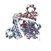
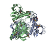
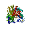
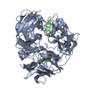
BIOMOLECULE: 1 THIS ENTRY CONTAINS THE CRYSTALLOGRAPHIC ASYMMETRIC UNIT WHICH CONSISTS OF 2 ... BIOMOLECULE: 1 THIS ENTRY CONTAINS THE CRYSTALLOGRAPHIC ASYMMETRIC UNIT WHICH CONSISTS OF 2 CHAIN(S). SEE REMARK 350 FOR INFORMATION ON GENERATING THE BIOLOGICAL MOLECULE(S). SIZE EXCLUSION CHROMATOGRAPHY WITH STATIC LIGHT SCATTERING SUPPORTS THE ASSIGNMENT OF A TETRAMER AS A BIOLOGICALLY SIGNIFICANT OLIGOMERIZATION STATE.
Type: MARMOSAIC 300 mm CCD / Detector: CCD / Date: Oct 19, 2006 / Details: Adjustable focusing mirrors in K-B geometry
Radiation
Monochromator: Si(111) Double Crystal Monochrometer / Protocol: SINGLE WAVELENGTH / Monochromatic (M) / Laue (L): M / Scattering type: x-ray
Radiation wavelength
Wavelength: 0.97922 Å / Relative weight: 1
Reflection
Resolution: 1.9→28.796 Å / Num. obs: 32500 / % possible obs: 99.6 % / Redundancy: 7.2 % / Biso Wilson estimate: 23.96 Å2 / Rmerge(I) obs: 0.193 / Rsym value: 0.193 / Net I/σ(I): 10
Reflection shell
Diffraction-ID: 1
Resolution (Å)
Redundancy (%)
Rmerge(I) obs
Mean I/σ(I) obs
Num. measured all
Num. unique all
Rsym value
% possible all
2-2.05
7.2
1.385
1.6
17122
2368
1.385
100
2.05-2.11
7.2
0.014
0.7
16667
2310
0.01064
100
2.11-2.17
7.3
0.014
0.8
16365
2251
0.861
100
2.17-2.24
7.2
0.014
1
15712
2182
0.74
99.9
2.24-2.31
7.2
0.014
1.1
15482
2136
0.639
99.9
2.31-2.39
7.2
0.014
1.3
14761
2041
0.548
99.9
2.39-2.48
7.2
0.014
1.6
14386
1988
0.451
99.8
2.48-2.58
7.3
0.014
1.8
13871
1911
0.41
99.8
2.58-2.7
7.2
0.014
2.3
13359
1852
0.319
99.8
2.7-2.83
7.2
0.014
2.9
12604
1748
0.251
99.7
2.83-2.98
7.2
0.014
3.7
12151
1682
0.2
99.6
2.98-3.16
7.2
0.014
4.6
11436
1589
0.157
99.5
3.16-3.38
7.2
0.014
5.2
10749
1490
0.133
99.4
3.38-3.65
7.1
0.014
5.9
9929
1397
0.114
99.3
3.65-4
7
0.014
6.7
9170
1301
0.1
99
4-4.47
7.1
0.014
7.9
8399
1177
0.081
98.8
4.47-5.16
7
0.014
9.3
7421
1053
0.07
98.9
5.16-6.32
7
0.014
8.5
6194
891
0.078
98.2
6.32-8.94
6.7
0.014
9.3
4831
718
0.07
98.2
8.94-28.8
6.1
0.014
8.4
2527
415
0.061
94.6
-
Phasing
Phasing
Method: SAD
-
Processing
Software
Name
Version
Classification
NB
MolProbity
3beta29
modelbuilding
SHELX
phasing
REFMAC
5.2.0005
refinement
SCALA
datascaling
PDB_EXTRACT
2
dataextraction
MOSFLM
datareduction
CCP4
(SCALA)
datascaling
SHELXD
phasing
autoSHARP
phasing
Refinement
Method to determine structure: SAD / Resolution: 2→28.796 Å / Cor.coef. Fo:Fc: 0.965 / Cor.coef. Fo:Fc free: 0.955 / SU B: 5.811 / SU ML: 0.082 / TLS residual ADP flag: LIKELY RESIDUAL / Cross valid method: THROUGHOUT / σ(F): 0 / ESU R: 0.12 / ESU R Free: 0.115 Stereochemistry target values: MAXIMUM LIKELIHOOD WITH PHASES Details: 1. HYDROGENS HAVE BEEN ADDED IN THE RIDING POSITIONS. 2. ATOM RECORD CONTAINS RESIDUAL B FACTORS ONLY. 3. A MET-INHIBITION PROTOCOL WAS USED FOR SELENOMETHIONINE INCORPORATION DURING PROTEIN ...Details: 1. HYDROGENS HAVE BEEN ADDED IN THE RIDING POSITIONS. 2. ATOM RECORD CONTAINS RESIDUAL B FACTORS ONLY. 3. A MET-INHIBITION PROTOCOL WAS USED FOR SELENOMETHIONINE INCORPORATION DURING PROTEIN EXPRESSION. THE OCCUPANCY OF THE SE ATOMS IN THE MSE RESIDUES WAS REDUCED TO 0.75 TO ACCOUNT FOR THE REDUCED SCATTERING POWER DUE TO PARTIAL S-MET INCORPORATION. 4. FIVE MPD MOLCULES FROM CRYSTALLIZATION SOLUTION ARE INCLUDED IN THE MODEL. 5. THERE IS SOME UNKNOWN DENSITY NEAR HIS64B, BUT THE EQUIVALENT DENSITY WAS NOT FOUND IN HIS64A.
Rfactor
Num. reflection
% reflection
Selection details
Rfree
0.187
1648
5.1 %
RANDOM
Rwork
0.155
-
-
-
obs
0.157
32500
99.29 %
-
Solvent computation
Ion probe radii: 0.8 Å / Shrinkage radii: 0.8 Å / VDW probe radii: 1.2 Å / Solvent model: BABINET MODEL WITH MASK
In the structure databanks used in Yorodumi, some data are registered as the other names, "COVID-19 virus" and "2019-nCoV". Here are the details of the virus and the list of structure data.
Jan 31, 2019. EMDB accession codes are about to change! (news from PDBe EMDB page)
EMDB accession codes are about to change! (news from PDBe EMDB page)
The allocation of 4 digits for EMDB accession codes will soon come to an end. Whilst these codes will remain in use, new EMDB accession codes will include an additional digit and will expand incrementally as the available range of codes is exhausted. The current 4-digit format prefixed with “EMD-” (i.e. EMD-XXXX) will advance to a 5-digit format (i.e. EMD-XXXXX), and so on. It is currently estimated that the 4-digit codes will be depleted around Spring 2019, at which point the 5-digit format will come into force.
The EM Navigator/Yorodumi systems omit the EMD- prefix.
Related info.:Q: What is EMD? / ID/Accession-code notation in Yorodumi/EM Navigator
Yorodumi is a browser for structure data from EMDB, PDB, SASBDB, etc.
This page is also the successor to EM Navigator detail page, and also detail information page/front-end page for Omokage search.
The word "yorodu" (or yorozu) is an old Japanese word meaning "ten thousand". "mi" (miru) is to see.
Related info.:EMDB / PDB / SASBDB / Comparison of 3 databanks / Yorodumi Search / Aug 31, 2016. New EM Navigator & Yorodumi / Yorodumi Papers / Jmol/JSmol / Function and homology information / Changes in new EM Navigator and Yorodumi
 Movie
Movie Controller

 Yorodumi
 Open data
 Basic information
 Components
 Keywords
 Function and homology information
 Jannaschia (bacteria)
 X-RAY DIFFRACTION /
  Authors
 Citation
 Structure visualization
 Downloads & links
 Other downloads
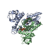


 PDBj
PDBj
 Assembly
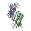


 Mass: 118.174 Da / Num. of mol.: 5 / Source method: obtained synthetically / Formula: C6H14O2 / Comment: precipitant*YM
 Mass: 18.015 Da / Num. of mol.: 221 / Source method: isolated from a natural source / Formula: H2O
 Sample preparation
 / Beamline: 23-ID-D / Wavelength: 0.97922
  Processing