 Movie
Movie Controller

[English] 日本語
 Yorodumi
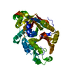
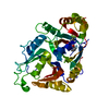
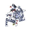
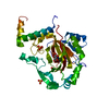
- PDB-2nsm: Crystal structure of the human carboxypeptidase N (Kininase I) ca... -
+ Open data

- Basic information
| Entry | Database: PDB / ID: 2nsm | ||||||
|---|---|---|---|---|---|---|---|
| Title | Crystal structure of the human carboxypeptidase N (Kininase I) catalytic domain | ||||||
 Components Components | Carboxypeptidase N catalytic chain | ||||||
 Keywords Keywords | HYDROLASE / caroxypeptidase / zinc peptidase / transthyretin-like domain / hormone processing / peptide modification | ||||||
| Function / homology |  Function and homology information Function and homology informationlysine carboxypeptidase / peptide metabolic process / bradykinin catabolic process / metallocarboxypeptidase activity / response to glucocorticoid / Regulation of Complement cascade / protein catabolic process / protein processing / extracellular space / extracellular region / zinc ion binding Similarity search - Function | ||||||
| Biological species |  Homo sapiens (human) Homo sapiens (human) | ||||||
| Method |  X-RAY DIFFRACTION / SYNCHROTRON / MOLECULAR REPLACEMENT / Resolution: 2.1 Å X-RAY DIFFRACTION / SYNCHROTRON / MOLECULAR REPLACEMENT / Resolution: 2.1 Å | ||||||
 Authors Authors | Keil, C. / Maskos, K. / Than, M. / Hoopes, J.T. / Huber, R. / Tan, F. / Deddish, P.A. / Erdoes, E.G. / Skidgel, R.A. / Bode, W. | ||||||
 Citation Citation | Journal: J.Mol.Biol. / Year: 2007 Title: Crystal structure of the human carboxypeptidase N (kininase I) catalytic domain Authors: Keil, C. / Maskos, K. / Than, M. / Hoopes, J.T. / Huber, R. / Tan, F. / Deddish, P.A. / Erdoes, E.G. / Skidgel, R.A. / Bode, W. | ||||||
| History |
|
- Structure visualization
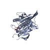
| Structure viewer | Molecule: MolmilJmol/JSmol |
|---|
- Downloads & links
-Download
| PDBx/mmCIF format | 2nsm.cif.gz | 103.2 KB | Display | PDBx/mmCIF format |
|---|---|---|---|---|
| PDB format | pdb2nsm.ent.gz | 76.4 KB | Display | PDB format |
| PDBx/mmJSON format | 2nsm.json.gz | Tree view | PDBx/mmJSON format | |
| Others |  Other downloads Other downloads |
-Validation report
| Summary document | 2nsm_validation.pdf.gz | 466 KB | Display | wwPDB validaton report |
|---|---|---|---|---|
| Full document | 2nsm_full_validation.pdf.gz | 469.5 KB | Display | |
| Data in XML | 2nsm_validation.xml.gz | 20.4 KB | Display | |
| Data in CIF | 2nsm_validation.cif.gz | 30.8 KB | Display | |
| Arichive directory | https://data.pdbj.org/pub/pdb/validation_reports/ns/2nsmftp://data.pdbj.org/pub/pdb/validation_reports/ns/2nsm | HTTPS FTP |
-Related structure data
| Related structure data |  1h8lS S: Starting model for refinement |
|---|---|
| Similar structure data |


-Links
 PDBj
PDBj
- Assembly
| Deposited unit | 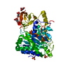
| ||||||||
|---|---|---|---|---|---|---|---|---|---|
| 1 |
| ||||||||
| Unit cell |
|
-Components
| #1: Protein | Mass: 50184.430 Da / Num. of mol.: 1 Source method: isolated from a genetically manipulated source Source: (gene. exp.) Homo sapiens (human) / Plasmid: pPIC9 / Production host:  Pichia pastoris (fungus) / Strain (production host): GS115 / References: UniProt: P15169, lysine carboxypeptidase Pichia pastoris (fungus) / Strain (production host): GS115 / References: UniProt: P15169, lysine carboxypeptidase | ||||||
|---|---|---|---|---|---|---|---|
| #2: Sugar |   Type: D-saccharide, beta linking / Mass: 221.208 Da / Num. of mol.: 3 Type: D-saccharide, beta linking / Mass: 221.208 Da / Num. of mol.: 3Source method: isolated from a genetically manipulated source Formula: C8H15NO6 #3: Chemical | ChemComp-SO4 /   Mass: 96.063 Da / Num. of mol.: 4 / Source method: obtained synthetically / Formula: SO4 Mass: 96.063 Da / Num. of mol.: 4 / Source method: obtained synthetically / Formula: SO4#4: Water | ChemComp-HOH / |  Mass: 18.015 Da / Num. of mol.: 370 / Source method: isolated from a natural source / Formula: H2O Mass: 18.015 Da / Num. of mol.: 370 / Source method: isolated from a natural source / Formula: H2OHas protein modification | Y | |
-Experimental details
-Experiment
| Experiment | Method: X-RAY DIFFRACTION / Number of used crystals: 1 |
|---|
- Sample preparation
| Crystal | Density Matthews: 3.55 Å3/Da / Density % sol: 65.38 % |
|---|---|
| Crystal grow | Temperature: 298 K / Method: vapor diffusion, sitting drop / pH: 7.5 Details: 0.1M Hepes, 2M Ammonium sulfate, 5mM EDTA, pH 7.5, VAPOR DIFFUSION, SITTING DROP, temperature 298K |
-Data collection
| Diffraction | Mean temperature: 100 K |
|---|---|
| Diffraction source | Source: SYNCHROTRON / Site: MPG/DESY, HAMBURG  / Beamline: BW6 / Wavelength: 1.05 Å / Beamline: BW6 / Wavelength: 1.05 Å |
| Detector | Type: MAR CCD 165 mm / Detector: CCD / Details: mirrors |
| Radiation | Monochromator: Si(111) double crystal, non dispersive / Protocol: SINGLE WAVELENGTH / Monochromatic (M) / Laue (L): M / Scattering type: x-ray |
| Radiation wavelength | Wavelength: 1.05 Å / Relative weight: 1 |
| Reflection | Resolution: 2.1→20 Å / Num. all: 41723 / Num. obs: 41723 / % possible obs: 99.4 % / Observed criterion σ(F): 4.37 / Observed criterion σ(I): 4.37 / Redundancy: 3.36 % / Biso Wilson estimate: 25 Å2 / Rmerge(I) obs: 0.068 / Rsym value: 0.068 / Net I/σ(I): 15.26 |
| Reflection shell | Resolution: 2.1→2.17 Å / Redundancy: 3.35 % / Rmerge(I) obs: 0.28 / Mean I/σ(I) obs: 4.37 / Num. unique all: 41723 / Rsym value: 0.28 / % possible all: 99.4 |
- Processing
| Software |
| ||||||||||||||||||||
|---|---|---|---|---|---|---|---|---|---|---|---|---|---|---|---|---|---|---|---|---|---|
| Refinement | Method to determine structure: MOLECULAR REPLACEMENT Starting model: PDB Entry 1H8L Resolution: 2.1→20 Å / σ(F): 4.37 / σ(I): 4.37 / Stereochemistry target values: Engh & Huber
| ||||||||||||||||||||
| Displacement parameters | Biso mean: 27.1 Å2 | ||||||||||||||||||||
| Refinement step | Cycle: LAST / Resolution: 2.1→20 Å
| ||||||||||||||||||||
| Refine LS restraints |
| ||||||||||||||||||||
| LS refinement shell | Resolution: 2.1→2.17 Å / Rfactor Rfree error: 0.012
|
