 Movie
Movie Controller


+ Open data

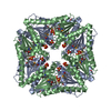
- Basic information
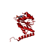
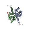
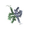
| Entry | Database: PDB / ID: 2nsj | ||||||
|---|---|---|---|---|---|---|---|
| Title | E. coli PurE H45Q mutant complexed with CAIR | ||||||
 Components Components | Phosphoribosylaminoimidazole carboxylase catalytic subunit | ||||||
 Keywords Keywords | LYASE / central three-layer alpha-beta-alpha sandwich / kinked C-terminal helix | ||||||
| Function / homology |  Function and homology information Function and homology information5-(carboxyamino)imidazole ribonucleotide mutase / 5-(carboxyamino)imidazole ribonucleotide mutase activity / 'de novo' IMP biosynthetic process / identical protein binding / cytosol Similarity search - Function | ||||||
| Biological species |  | ||||||
| Method |  X-RAY DIFFRACTION / SYNCHROTRON / MOLECULAR REPLACEMENT / Resolution: 2.31 Å X-RAY DIFFRACTION / SYNCHROTRON / MOLECULAR REPLACEMENT / Resolution: 2.31 Å | ||||||
 Authors Authors | Ealick, S.E. / Morar, M. | ||||||
 Citation Citation | Journal: Biochemistry / Year: 2007 Title: N(5)-CAIR Mutase: Role of a CO(2) Binding Site and Substrate Movement in Catalysis. Authors: Hoskins, A.A. / Morar, M. / Kappock, T.J. / Mathews, I.I. / Zaugg, J.B. / Barder, T.E. / Peng, P. / Okamoto, A. / Ealick, S.E. / Stubbe, J. | ||||||
| History |
|
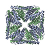
- Structure visualization
| Structure viewer | Molecule: MolmilJmol/JSmol |
|---|
- Downloads & links
-Download
| PDBx/mmCIF format | 2nsj.cif.gz | 45.7 KB | Display | PDBx/mmCIF format |
|---|---|---|---|---|
| PDB format | pdb2nsj.ent.gz | 30.9 KB | Display | PDB format |
| PDBx/mmJSON format | 2nsj.json.gz | Tree view | PDBx/mmJSON format | |
| Others |  Other downloads Other downloads |
-Validation report
| Arichive directory | https://data.pdbj.org/pub/pdb/validation_reports/ns/2nsjftp://data.pdbj.org/pub/pdb/validation_reports/ns/2nsj | HTTPS FTP |
|---|
-Related structure data
| Related structure data | 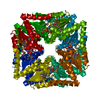 2ateSC  2nshC  2nslC S: Starting model for refinement C: citing same article ( |
|---|---|
| Similar structure data |

-Links
 PDBj
PDBj- Assembly
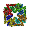
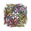
| Deposited unit | 
| ||||||||
|---|---|---|---|---|---|---|---|---|---|
| 1 | x 8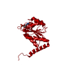
| ||||||||
| Unit cell |
|
-Components
| #1: Protein | Mass: 17789.252 Da / Num. of mol.: 1 / Mutation: H45Q Source method: isolated from a genetically manipulated source Source: (gene. exp.) References: UniProt: P0AG18, phosphoribosylaminoimidazole carboxylase |
|---|---|
| #2: Chemical | ChemComp-C2R /   Mass: 339.196 Da / Num. of mol.: 1 / Source method: obtained synthetically / Formula: C9H14N3O9P Mass: 339.196 Da / Num. of mol.: 1 / Source method: obtained synthetically / Formula: C9H14N3O9P |
| #3: Water | ChemComp-HOH /  Mass: 18.015 Da / Num. of mol.: 62 / Source method: isolated from a natural source / Formula: H2O Mass: 18.015 Da / Num. of mol.: 62 / Source method: isolated from a natural source / Formula: H2O |
-Experimental details
-Experiment
| Experiment | Method: X-RAY DIFFRACTION / Number of used crystals: 1 |
|---|
- Sample preparation
| Crystal | Density Matthews: 2.15 Å3/Da / Density % sol: 42.71 % |
|---|---|
| Crystal grow | Temperature: 298 K / Method: vapor diffusion, hanging drop / pH: 8 Details: 25% PEG400, 0.2M magnesium chloride, 0.1M Tris, pH 8.0, VAPOR DIFFUSION, HANGING DROP, temperature 298K |
-Data collection
| Diffraction | Mean temperature: 100 K |
|---|---|
| Diffraction source | Source: SYNCHROTRON / Site: APS  / Beamline: 24-ID-C / Wavelength: 0.9795 / Beamline: 24-ID-C / Wavelength: 0.9795 |
| Detector | Type: ADSC QUANTUM 315 / Detector: CCD / Date: Jun 5, 2006 |
| Radiation | Protocol: SINGLE WAVELENGTH / Monochromatic (M) / Laue (L): M / Scattering type: x-ray |
| Radiation wavelength | Wavelength: 0.9795 Å / Relative weight: 1 |
| Reflection | Resolution: 2.31→39.34 Å / Num. all: 8526 / Num. obs: 6763 / % possible obs: 95.8 % / Observed criterion σ(F): 0 / Observed criterion σ(I): 0 / Biso Wilson estimate: 30 Å2 |
| Reflection shell | Resolution: 2.31→2.44 Å / % possible all: 86.4 |
- Processing
| Software |
| ||||||||||||||||||||||||||||||||||||||||||||||||||||||||||||||||||||||||||||||||
|---|---|---|---|---|---|---|---|---|---|---|---|---|---|---|---|---|---|---|---|---|---|---|---|---|---|---|---|---|---|---|---|---|---|---|---|---|---|---|---|---|---|---|---|---|---|---|---|---|---|---|---|---|---|---|---|---|---|---|---|---|---|---|---|---|---|---|---|---|---|---|---|---|---|---|---|---|---|---|---|---|---|
| Refinement | Method to determine structure: MOLECULAR REPLACEMENT Starting model: PDB ENTRY 2ATE Resolution: 2.31→39.34 Å / Rfactor Rfree error: 0.009 / Data cutoff high absF: 345703.32 / Data cutoff low absF: 0 / Isotropic thermal model: RESTRAINED / Cross valid method: THROUGHOUT / σ(F): 0
| ||||||||||||||||||||||||||||||||||||||||||||||||||||||||||||||||||||||||||||||||
| Solvent computation | Solvent model: FLAT MODEL / Bsol: 41.7116 Å2 / ksol: 0.358729 e/Å3 | ||||||||||||||||||||||||||||||||||||||||||||||||||||||||||||||||||||||||||||||||
| Displacement parameters | Biso mean: 40.2 Å2
| ||||||||||||||||||||||||||||||||||||||||||||||||||||||||||||||||||||||||||||||||
| Refine analyze |
| ||||||||||||||||||||||||||||||||||||||||||||||||||||||||||||||||||||||||||||||||
| Refinement step | Cycle: LAST / Resolution: 2.31→39.34 Å
| ||||||||||||||||||||||||||||||||||||||||||||||||||||||||||||||||||||||||||||||||
| Refine LS restraints |
| ||||||||||||||||||||||||||||||||||||||||||||||||||||||||||||||||||||||||||||||||
| LS refinement shell | Resolution: 2.31→2.44 Å / Rfactor Rfree error: 0.031 / Total num. of bins used: 6
| ||||||||||||||||||||||||||||||||||||||||||||||||||||||||||||||||||||||||||||||||
| Xplor file |
|
