 Movie
Movie Controller

[English] 日本語
 Yorodumi
- PDB-2mwg: Full-Length Solution Structure Of YtvA, a LOV-Photoreceptor Prote... -
+ Open data

- Basic information
| Entry | Database: PDB / ID: 2mwg | ||||||
|---|---|---|---|---|---|---|---|
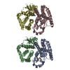
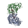
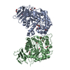
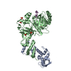
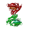
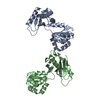
| Title | Full-Length Solution Structure Of YtvA, a LOV-Photoreceptor Protein and Regulator of Bacterial Stress Response | ||||||
 Components Components | Blue-light photoreceptor | ||||||
 Keywords Keywords | PROTEIN BINDING / Photoreceptor / LOV/PAS / Stressosome / Rsb | ||||||
| Function / homology |  Function and homology information Function and homology information | ||||||
| Biological species |  | ||||||
| Method | SOLUTION NMR / monte-carlo simulated annealing using torsion angle dynamics, simulated annealing. | ||||||
 Authors Authors | Jurk, M. / Bardiaux, B. / Schmieder, P. | ||||||
 Citation Citation | Journal: To be Published Title: Solution Structure of YtvA from Bacillus subtilis Provides Insight into Activation Mechanism and Regulation of Bacterial Stress Response. Authors: Jurk, M. / Dorn, M. / Reichenwallner, J. / Bardiaux, B. / Hinderberger, D. / Schmieder, P. | ||||||
| History |
|
- Structure visualization
| Structure viewer | Molecule: MolmilJmol/JSmol |
|---|
- Downloads & links
-Download
| PDBx/mmCIF format | 2mwg.cif.gz | 1.6 MB | Display | PDBx/mmCIF format |
|---|---|---|---|---|
| PDB format | pdb2mwg.ent.gz | 1.4 MB | Display | PDB format |
| PDBx/mmJSON format | 2mwg.json.gz | Tree view | PDBx/mmJSON format | |
| Others |  Other downloads Other downloads |
-Validation report
| Arichive directory | https://data.pdbj.org/pub/pdb/validation_reports/mw/2mwgftp://data.pdbj.org/pub/pdb/validation_reports/mw/2mwg | HTTPS FTP |
|---|
-Related structure data
| Similar structure data | |
|---|---|
| Other databases |
|

-Links
 PDBj
PDBj


- Assembly
| Deposited unit | 
| |||||||||
|---|---|---|---|---|---|---|---|---|---|---|
| 1 |
| |||||||||
| NMR ensembles |
|
-Components
| #1: Protein | Mass: 29148.361 Da / Num. of mol.: 2 / Fragment: UNP residues 2-261 Source method: isolated from a genetically manipulated source Source: (gene. exp.) #2: Chemical |   Mass: 456.344 Da / Num. of mol.: 2 / Source method: obtained synthetically / Formula: C17H21N4O9P Mass: 456.344 Da / Num. of mol.: 2 / Source method: obtained synthetically / Formula: C17H21N4O9P |
|---|
-Experimental details
-Experiment
| Experiment | Method: SOLUTION NMR | ||||||||||||||||||||
|---|---|---|---|---|---|---|---|---|---|---|---|---|---|---|---|---|---|---|---|---|---|
| NMR experiment |
|
- Sample preparation
| Details |
| ||||||||||||||||||||||||||||||||||||||||||||||||||||||||||||||||||||||||||||||||||||||||||||||||
|---|---|---|---|---|---|---|---|---|---|---|---|---|---|---|---|---|---|---|---|---|---|---|---|---|---|---|---|---|---|---|---|---|---|---|---|---|---|---|---|---|---|---|---|---|---|---|---|---|---|---|---|---|---|---|---|---|---|---|---|---|---|---|---|---|---|---|---|---|---|---|---|---|---|---|---|---|---|---|---|---|---|---|---|---|---|---|---|---|---|---|---|---|---|---|---|---|---|
| Sample |
| ||||||||||||||||||||||||||||||||||||||||||||||||||||||||||||||||||||||||||||||||||||||||||||||||
| Sample conditions | Ionic strength: 70 / pH: 6.5 / Pressure: ambient / Temperature: 300 K |
-NMR measurement
| NMR spectrometer | Type: Bruker Avance / Manufacturer: Bruker / Model: AVANCE / Field strength: 600 MHz |
|---|
- Processing
| NMR software |
| ||||||||||||||||||||||||||||||||||||||||
|---|---|---|---|---|---|---|---|---|---|---|---|---|---|---|---|---|---|---|---|---|---|---|---|---|---|---|---|---|---|---|---|---|---|---|---|---|---|---|---|---|---|
| Refinement | Method: monte-carlo simulated annealing using torsion angle dynamics, simulated annealing. Software ordinal: 1 Details: OWING TO THE RELATIVELY LOW NUMBER OF RESTRAINTS TO DETERMINE THE STRUCTURE AB INITIO, STRUCTURES OF THE FOUR MAJOR SEGMENTS WERE CALCULATED USING CS-ROSETTA. 12,000 AND 2,000 STRUCTURES ...Details: OWING TO THE RELATIVELY LOW NUMBER OF RESTRAINTS TO DETERMINE THE STRUCTURE AB INITIO, STRUCTURES OF THE FOUR MAJOR SEGMENTS WERE CALCULATED USING CS-ROSETTA. 12,000 AND 2,000 STRUCTURES WERE CALCULATED FOR LOV OR STAS AND NCAP OR JA, RESPECTIVELY. THE LOWEST ENERGY STRUCTURE WAS FURTHER USED. THE STRUCTURES OF THE FOUR SEGMENTS OBTAINED BY CS-ROSETTA WERE REFINED AGAINST THE RDCS USING LOW TEMPERATURE SA. 100 STRUCTURES WERE CALCULATED AND THE CLOSEST TO AN AVERAGE OF THE 10 LOWEST ENERGY STRUCTURES WAS USED IN THE NEXT STEP. ALL FOUR SEGMENTS (NCAP, LOV, JA, STAS) WERE COMBINED TO A DIMERIC FULL-LENGTH STRUCTURE. THE YF1 STRUCTURE (PDB 4GCZ) WAS USED AS A TEMPLATE FOR THE ALIGNMENT OF LOV AND NCAP IN THE DIMER. ALL FOUR SEGMENTS WERE TREATED AS RIGID BODIES IN A HIGH TEMPERATURE SIMULATED ANNEALING. 100 STRUCTURES WERE CALCULATED AND THE CLOSEST TO AN AVERAGE OF THE LOWEST 10 WAS USED IN THE LAST REFINEMENT STEP. THE FINAL STRUCTURAL ENSEMBLE WAS OBTAINED USING THE SAME REFINEMENT PROTOCOLS AS IN STEP 2. 100 STRUCTURES WERE CALCULATED AND THE LOWEST 10 ARE SUBMITTED. | ||||||||||||||||||||||||||||||||||||||||
| NMR constraints | NOE constraints total: 1192 / NOE intraresidue total count: 58 / NOE long range total count: 294 / NOE medium range total count: 380 / NOE sequential total count: 452 / Protein chi angle constraints total count: 0 / Protein other angle constraints total count: 0 / Protein phi angle constraints total count: 412 / Protein psi angle constraints total count: 402 | ||||||||||||||||||||||||||||||||||||||||
| NMR representative | Selection criteria: closest to the average | ||||||||||||||||||||||||||||||||||||||||
| NMR ensemble | Conformer selection criteria: structures with the lowest energy Conformers calculated total number: 100 / Conformers submitted total number: 10 |
 X-PLOR NIH