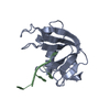
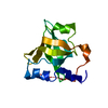
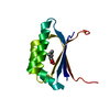
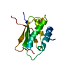
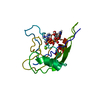
- PDB-2m7s: NMR structure of RNA recognition motif 2 (RRM2) of Homo sapiens s... -
+
Open data
ID or keywords:
Loading...
-
Basic information
Entry
Database: PDB / ID: 2m7s
Title
NMR structure of RNA recognition motif 2 (RRM2) of Homo sapiens splicing factor, arginine/serine-rich 1
Components
Serine/arginine-rich splicing factor 1
Keywords
RNA BINDING PROTEIN / RNA Recognition Motif / Structural Genomics / PSI-Biology / Joint Center for Structural Genomics / JCSG / Partnership for T-Cell Biology / TCELL
Function / homology
Function and homology information
protein localization to P-body / DNA topoisomerase binding / RS domain binding / protein kinase B binding / interleukin-17-mediated signaling pathway / mRNA splice site recognition / alternative mRNA splicing, via spliceosome / signal transduction involved in regulation of gene expression / mRNA 3'-end processing / Transport of Mature mRNA derived from an Intron-Containing Transcript ...protein localization to P-body / DNA topoisomerase binding / RS domain binding / protein kinase B binding / interleukin-17-mediated signaling pathway / mRNA splice site recognition / alternative mRNA splicing, via spliceosome / signal transduction involved in regulation of gene expression / mRNA 3'-end processing / Transport of Mature mRNA derived from an Intron-Containing Transcript / RNA Polymerase II Transcription Termination / mRNA stabilization / regulation of alternative mRNA splicing, via spliceosome / mRNA 5'-splice site recognition / regulation of RNA splicing / mRNA Splicing - Minor Pathway / oligodendrocyte differentiation / Processing of Capped Intron-Containing Pre-mRNA / mRNA transport / protein localization to nucleus / catalytic step 2 spliceosome / cardiac muscle contraction / mRNA Splicing - Major Pathway / liver regeneration / positive regulation of RNA splicing / mRNA splicing, via spliceosome / mRNA processing / nuclear envelope / in utero embryonic development / nuclear speck / mRNA binding / RNA binding / nucleoplasm / nucleus / cytoplasm Similarity search - Function
NOE constraints total: 1414 / NOE intraresidue total count: 317 / NOE long range total count: 479 / NOE medium range total count: 204 / NOE sequential total count: 394
NMR representative
Selection criteria: closest to the average
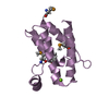
NMR ensemble
Conformer selection criteria: target function / Conformers calculated total number: 80 / Conformers submitted total number: 20
+
About Yorodumi
-
News
-
Feb 9, 2022. New format data for meta-information of EMDB entries
New format data for meta-information of EMDB entries
Version 3 of the EMDB header file is now the official format.
The previous official version 1.9 will be removed from the archive.
In the structure databanks used in Yorodumi, some data are registered as the other names, "COVID-19 virus" and "2019-nCoV". Here are the details of the virus and the list of structure data.
Jan 31, 2019. EMDB accession codes are about to change! (news from PDBe EMDB page)
EMDB accession codes are about to change! (news from PDBe EMDB page)
The allocation of 4 digits for EMDB accession codes will soon come to an end. Whilst these codes will remain in use, new EMDB accession codes will include an additional digit and will expand incrementally as the available range of codes is exhausted. The current 4-digit format prefixed with “EMD-” (i.e. EMD-XXXX) will advance to a 5-digit format (i.e. EMD-XXXXX), and so on. It is currently estimated that the 4-digit codes will be depleted around Spring 2019, at which point the 5-digit format will come into force.
The EM Navigator/Yorodumi systems omit the EMD- prefix.
Related info.:Q: What is EMD? / ID/Accession-code notation in Yorodumi/EM Navigator
Yorodumi is a browser for structure data from EMDB, PDB, SASBDB, etc.
This page is also the successor to EM Navigator detail page, and also detail information page/front-end page for Omokage search.
The word "yorodu" (or yorozu) is an old Japanese word meaning "ten thousand". "mi" (miru) is to see.
Related info.:EMDB / PDB / SASBDB / Comparison of 3 databanks / Yorodumi Search / Aug 31, 2016. New EM Navigator & Yorodumi / Yorodumi Papers / Jmol/JSmol / Function and homology information / Changes in new EM Navigator and Yorodumi
 Movie
Movie Controller

 Yorodumi
 Open data
 Basic information
 Components
 Keywords
 Function and homology information
 Homo sapiens (human)
 Authors
 Citation
 Structure visualization
 Downloads & links
 Other downloads


 PDBj
PDBj


 Assembly
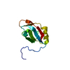

 HSQC
HSQC Sample preparation
 Processing