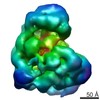
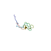
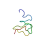
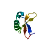
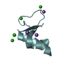
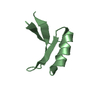
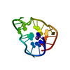
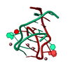
Journal: Nat Struct Mol Biol / Year: 2013 Title: Electron microscopy structure of human APC/C(CDH1)-EMI1 reveals multimodal mechanism of E3 ligase shutdown. Authors: Jeremiah J Frye / Nicholas G Brown / Georg Petzold / Edmond R Watson / Christy R R Grace / Amanda Nourse / Marc A Jarvis / Richard W Kriwacki / Jan-Michael Peters / Holger Stark / Brenda A Schulman / Abstract: The anaphase-promoting complex/cyclosome (APC/C) is a ~1.5-MDa multiprotein E3 ligase enzyme that regulates cell division by promoting timely ubiquitin-mediated proteolysis of key cell-cycle ...The anaphase-promoting complex/cyclosome (APC/C) is a ~1.5-MDa multiprotein E3 ligase enzyme that regulates cell division by promoting timely ubiquitin-mediated proteolysis of key cell-cycle regulatory proteins. Inhibition of human APC/C(CDH1) during interphase by early mitotic inhibitor 1 (EMI1) is essential for accurate coordination of DNA synthesis and mitosis. Here, we report a hybrid structural approach involving NMR, electron microscopy and enzymology, which reveal that EMI1's 143-residue C-terminal domain inhibits multiple APC/C(CDH1) functions. The intrinsically disordered D-box, linker and tail elements, together with a structured zinc-binding domain, bind distinct regions of APC/C(CDH1) to synergistically both block the substrate-binding site and inhibit ubiquitin-chain elongation. The functional importance of intrinsic structural disorder is explained by enabling a small inhibitory domain to bind multiple sites to shut down various functions of a 'molecular machine' nearly 100 times its size.
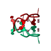
NOE constraints total: 931 / NOE intraresidue total count: 211 / NOE long range total count: 256 / NOE medium range total count: 0 / NOE sequential total count: 83 / Protein chi angle constraints total count: 0 / Protein other angle constraints total count: 0 / Protein phi angle constraints total count: 19 / Protein psi angle constraints total count: 19
NMR representative
Selection criteria: lowest energy
NMR ensemble
Average torsion angle constraint violation: 0.1997 ° / Conformer selection criteria: target function / Conformers calculated total number: 100 / Conformers submitted total number: 20 / Maximum lower distance constraint violation: 0.0093 Å / Maximum torsion angle constraint violation: 0.86 ° / Maximum upper distance constraint violation: 0.21 Å / Torsion angle constraint violation method: Talos program
NMR ensemble rms
Distance rms dev: 0.046 Å / Distance rms dev error: 0.004 Å
+
About Yorodumi
-
News
-
Feb 9, 2022. New format data for meta-information of EMDB entries
New format data for meta-information of EMDB entries
Version 3 of the EMDB header file is now the official format.
The previous official version 1.9 will be removed from the archive.
In the structure databanks used in Yorodumi, some data are registered as the other names, "COVID-19 virus" and "2019-nCoV". Here are the details of the virus and the list of structure data.
Jan 31, 2019. EMDB accession codes are about to change! (news from PDBe EMDB page)
EMDB accession codes are about to change! (news from PDBe EMDB page)
The allocation of 4 digits for EMDB accession codes will soon come to an end. Whilst these codes will remain in use, new EMDB accession codes will include an additional digit and will expand incrementally as the available range of codes is exhausted. The current 4-digit format prefixed with “EMD-” (i.e. EMD-XXXX) will advance to a 5-digit format (i.e. EMD-XXXXX), and so on. It is currently estimated that the 4-digit codes will be depleted around Spring 2019, at which point the 5-digit format will come into force.
The EM Navigator/Yorodumi systems omit the EMD- prefix.
Related info.:Q: What is EMD? / ID/Accession-code notation in Yorodumi/EM Navigator
Yorodumi is a browser for structure data from EMDB, PDB, SASBDB, etc.
This page is also the successor to EM Navigator detail page, and also detail information page/front-end page for Omokage search.
The word "yorodu" (or yorozu) is an old Japanese word meaning "ten thousand". "mi" (miru) is to see.
Related info.:EMDB / PDB / SASBDB / Comparison of 3 databanks / Yorodumi Search / Aug 31, 2016. New EM Navigator & Yorodumi / Yorodumi Papers / Jmol/JSmol / Function and homology information / Changes in new EM Navigator and Yorodumi
 Movie
Movie Controller


 Open data
 Basic information
 Components
 Keywords
 Function and homology information
 Homo sapiens (human)
 Authors
 Citation
 Structure visualization
 Downloads & links
 Other downloads
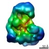


 PDBj
PDBj


 Assembly
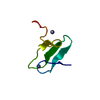


 Mass: 65.409 Da / Num. of mol.: 2 / Source method: obtained synthetically / Formula: Zn
 HSQC
HSQC Sample preparation
 Processing