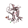
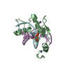
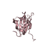
登録情報 データベース : PDB / ID : 2kohタイトル NMR structure of mouse Par3-PDZ3 in complex with VE-Cadherin C-terminus Cadherin-5 Partitioning defective 3 homolog キーワード / / / / / / / / / / / / / / / / / / / 機能・相同性 分子機能 ドメイン・相同性 構成要素
/ / / / / / / / / / / / / / / / / / / / / / / / / / / / / / / / / / / / / / / / / / / / / / / / / / / / / / / / / / / / / / / / / / / / / / / / / / / / / / / / / / / / / / / / / / / / / / / / / / / / / / / / / / / / / / / / / / / / / / / / / / / / / / / / / / / / / / 生物種 Mus musculus (ハツカネズミ)手法 / Model details lowest energy, model 1 データ登録者 Volkman, B.F. / Tyler, R.C. / Peterson, F.C. ジャーナル : Biochemistry / 年 : 2010タイトル : Distal interactions within the par3-VE-cadherin complex.著者 : Tyler, R.C. / Peterson, F.C. / Volkman, B.F. 履歴 登録 2009年9月22日 登録サイト / 処理サイト 改定 1.0 2010年2月9日 Provider / タイプ 改定 1.1 2011年7月13日 Group 改定 1.2 2022年3月16日 Group / Database references / Derived calculationsカテゴリ database_2 / pdbx_nmr_software ... database_2 / pdbx_nmr_software / pdbx_nmr_spectrometer / pdbx_struct_assembly / pdbx_struct_oper_list / struct_ref_seq_dif Item _database_2.pdbx_DOI / _database_2.pdbx_database_accession ... _database_2.pdbx_DOI / _database_2.pdbx_database_accession / _pdbx_nmr_software.name / _pdbx_nmr_spectrometer.model / _struct_ref_seq_dif.details 改定 1.3 2024年5月22日 Group / カテゴリ / chem_comp_bond
すべて表示 表示を減らす
 ムービー
ムービー コントローラー


 データを開く
 基本情報
基本情報 要素
要素 キーワード
キーワード 機能・相同性情報
 データ登録者
 引用
引用 構造の表示
構造の表示 ダウンロードとリンク
 その他のダウンロード
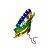


 PDBj
PDBj
 集合体
集合体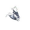

 試料調製
試料調製 解析
解析 Xplor-NIH