 Movie
Movie Controller


+ Open data

- Basic information
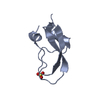
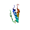
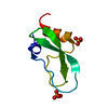
| Entry | Database: PDB / ID: 2knt | ||||||
|---|---|---|---|---|---|---|---|
| Title | THE 1.2 ANGSTROM STRUCTURE OF KUNITZ TYPE DOMAIN C5 | ||||||
 Components Components | COLLAGEN | ||||||
 Keywords Keywords | KUNITZ INHIBITOR / EXTRACELLULAR MATRIX / CONNECTIVE TISSUE | ||||||
| Function / homology |  Function and homology information Function and homology informationcollagen type VI trimer / Collagen chain trimerization / extracellular matrix structural constituent conferring tensile strength / Collagen biosynthesis and modifying enzymes / Signaling by PDGF / NCAM1 interactions / Assembly of collagen fibrils and other multimeric structures / muscle organ development / Collagen degradation / ECM proteoglycans ...collagen type VI trimer / Collagen chain trimerization / extracellular matrix structural constituent conferring tensile strength / Collagen biosynthesis and modifying enzymes / Signaling by PDGF / NCAM1 interactions / Assembly of collagen fibrils and other multimeric structures / muscle organ development / Collagen degradation / ECM proteoglycans / Integrin cell surface interactions / response to glucose / response to UV / phosphatidylinositol 3-kinase/protein kinase B signal transduction / serine-type endopeptidase inhibitor activity / sarcolemma / neuron apoptotic process / extracellular vesicle / extracellular matrix / cell adhesion / endoplasmic reticulum lumen / : / extracellular exosome / extracellular region Similarity search - Function | ||||||
| Biological species |  Homo sapiens (human) Homo sapiens (human) | ||||||
| Method |  X-RAY DIFFRACTION / SYNCHROTRON / MOLECULAR REPLACEMENT / Resolution: 1.2 Å X-RAY DIFFRACTION / SYNCHROTRON / MOLECULAR REPLACEMENT / Resolution: 1.2 Å | ||||||
 Authors Authors | Merigeau, K. / Arnoux, B. / Ducruix, A. | ||||||
 Citation Citation | Journal: Acta Crystallogr.,Sect.D / Year: 1998 Title: 1.2 A refinement of the Kunitz-type domain from the alpha3 chain of human type VI collagen. Authors: Merigeau, K. / Arnoux, B. / Perahia, D. / Norris, K. / Norris, F. / Ducruix, A. | ||||||
| History |
|
- Structure visualization
| Structure viewer | Molecule: MolmilJmol/JSmol |
|---|
- Downloads & links
-Download
| PDBx/mmCIF format | 2knt.cif.gz | 27.4 KB | Display | PDBx/mmCIF format |
|---|---|---|---|---|
| PDB format | pdb2knt.ent.gz | 15.1 KB | Display | PDB format |
| PDBx/mmJSON format | 2knt.json.gz | Tree view | PDBx/mmJSON format | |
| Others |  Other downloads Other downloads |
-Validation report
| Arichive directory | https://data.pdbj.org/pub/pdb/validation_reports/kn/2kntftp://data.pdbj.org/pub/pdb/validation_reports/kn/2knt | HTTPS FTP |
|---|
-Related structure data
| Related structure data |  1aapS S: Starting model for refinement |
|---|---|
| Similar structure data |


-Links
 PDBj
PDBj


- Assembly
| Deposited unit | 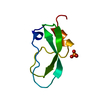
| ||||||||
|---|---|---|---|---|---|---|---|---|---|
| 1 |
| ||||||||
| Unit cell |
|
-Components
| #1: Protein | Mass: 6639.508 Da / Num. of mol.: 1 / Fragment: DOMAIN C5, C-TERMINUS OF TYPE VI COLLAGEN Source method: isolated from a genetically manipulated source Details: KUNITZ-TYPE / Source: (gene. exp.) Homo sapiens (human) / Production host:  |
|---|---|
| #2: Chemical | ChemComp-PO4 /   Mass: 94.971 Da / Num. of mol.: 1 / Source method: obtained synthetically / Formula: PO4 Mass: 94.971 Da / Num. of mol.: 1 / Source method: obtained synthetically / Formula: PO4 |
| #3: Water | ChemComp-HOH /  Mass: 18.015 Da / Num. of mol.: 50 / Source method: isolated from a natural source / Formula: H2O Mass: 18.015 Da / Num. of mol.: 50 / Source method: isolated from a natural source / Formula: H2O |
| Has protein modification | Y |
-Experimental details
-Experiment
| Experiment | Method: X-RAY DIFFRACTION / Number of used crystals: 2 |
|---|
- Sample preparation
| Crystal | Density Matthews: 1.99 Å3/Da / Density % sol: 39 % | ||||||||||||||||||||||||||||||||||||
|---|---|---|---|---|---|---|---|---|---|---|---|---|---|---|---|---|---|---|---|---|---|---|---|---|---|---|---|---|---|---|---|---|---|---|---|---|---|
| Crystal grow | pH: 3.3 Details: 0.2M LI2SO4, 0.1M CITRIC ACID, 0.074M NA2HPO4, 1.6M (NH4)2SO4, 10MG/ML, PH3.3 | ||||||||||||||||||||||||||||||||||||
| Crystal grow | *PLUS Temperature: 4 ℃ / pH: 3 / Method: vapor diffusion, sitting drop / Details: Arnoux, B., (1995) J. Mol. Biol., 246, 609. | ||||||||||||||||||||||||||||||||||||
| Components of the solutions | *PLUS
|
-Data collection
| Diffraction | Mean temperature: 291 K |
|---|---|
| Diffraction source | Source: SYNCHROTRON / Site: LURE  / Beamline: DW32 / Wavelength: 0.901 / Beamline: DW32 / Wavelength: 0.901 |
| Detector | Type: MARRESEARCH / Detector: IMAGE PLATE / Date: Dec 14, 1995 |
| Radiation | Monochromator: YES / Monochromatic (M) / Laue (L): M / Scattering type: x-ray |
| Radiation wavelength | Wavelength: 0.901 Å / Relative weight: 1 |
| Reflection | Resolution: 1.18→20.4 Å / Num. obs: 16657 / % possible obs: 96 % / Observed criterion σ(I): 0 / Redundancy: 4.9 % / Rmerge(I) obs: 0.055 / Net I/σ(I): 46.5 |
| Reflection shell | Resolution: 1.18→1.21 Å / Redundancy: 2.4 % / Mean I/σ(I) obs: 1.09 / Rsym value: 0.75 / % possible all: 52.5 |
| Reflection | *PLUS Num. measured all: 84000 |
| Reflection shell | *PLUS % possible obs: 61 % |
- Processing
| Software |
| |||||||||||||||||||||||||||||||||
|---|---|---|---|---|---|---|---|---|---|---|---|---|---|---|---|---|---|---|---|---|---|---|---|---|---|---|---|---|---|---|---|---|---|---|
| Refinement | Method to determine structure: MOLECULAR REPLACEMENT Starting model: PDB ENTRY 1AAP Resolution: 1.2→7 Å / Num. restraintsaints: 5876 / Cross valid method: FREE R-VALUE / σ(F): 0 / Stereochemistry target values: ENGH AND HUBER
| |||||||||||||||||||||||||||||||||
| Solvent computation | Solvent model: BABINET | |||||||||||||||||||||||||||||||||
| Refine analyze | Num. disordered residues: 5 / Occupancy sum hydrogen: 630 / Occupancy sum non hydrogen: 476 | |||||||||||||||||||||||||||||||||
| Refinement step | Cycle: LAST / Resolution: 1.2→7 Å
| |||||||||||||||||||||||||||||||||
| Refine LS restraints |
| |||||||||||||||||||||||||||||||||
| Software | *PLUS Name: SHELXL-93 / Classification: refinement | |||||||||||||||||||||||||||||||||
| Refinement | *PLUS Num. reflection obs: 10762 | |||||||||||||||||||||||||||||||||
| Solvent computation | *PLUS | |||||||||||||||||||||||||||||||||
| Displacement parameters | *PLUS |
