IgG binding / error-prone translesion synthesis / translesion synthesis / Translesion synthesis by POLI / Termination of translesion DNA synthesis / cytoplasmic ribonucleoprotein granule / DNA-directed DNA polymerase / damaged DNA binding / DNA-directed DNA polymerase activity / DNA replication ...IgG binding / error-prone translesion synthesis / translesion synthesis / Translesion synthesis by POLI / Termination of translesion DNA synthesis / cytoplasmic ribonucleoprotein granule / DNA-directed DNA polymerase / damaged DNA binding / DNA-directed DNA polymerase activity / DNA replication / nuclear speck / DNA repair / nucleoplasm / metal ion binding Similarity search - Function
HUWE1/Rev1, ubiquitin binding region / Ubiquitin binding region / : / DNA polymerase-iota, thumb domain / IgG-binding B / B domain / M protein-type anchor domain / Ubiquitin-binding motif (UBM) domain profile. / GA-like domain / GA-like domain ...HUWE1/Rev1, ubiquitin binding region / Ubiquitin binding region / : / DNA polymerase-iota, thumb domain / IgG-binding B / B domain / M protein-type anchor domain / Ubiquitin-binding motif (UBM) domain profile. / GA-like domain / GA-like domain / Immunoglobulin/albumin-binding domain superfamily / YSIRK Gram-positive signal peptide / DNA polymerase, Y-family, little finger domain / impB/mucB/samB family C-terminal domain / UmuC domain / DNA polymerase, Y-family, little finger domain superfamily / impB/mucB/samB family / UmuC domain profile. / LPXTG cell wall anchor motif / Gram-positive cocci surface proteins LPxTG motif profile. / LPXTG cell wall anchor domain / Reverse transcriptase/Diguanylate cyclase domain / DNA/RNA polymerase superfamily Similarity search - Domain/homology
ImmunoglobulinG-bindingproteinG, DNApolymeraseiota / IgG-binding protein G / RAD30 homolog B / Eta2
Mass: 12348.556 Da / Num. of mol.: 1 / Fragment: UNP residues 304-357, 676-715 Source method: isolated from a genetically manipulated source Source: (gene. exp.) Streptococcus sp., Homo sapiens / Gene: spg, POLI, RAD30B / Production host: Escherichia coli (E. coli) References: UniProt: P19909, UniProt: Q9UNA4, DNA-directed DNA polymerase
Sequence details
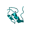
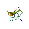
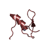
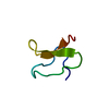
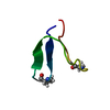
ACCORDING TO THE AUTHORS THE N-TERMINAL PART IS A GB1 TAG USED FOR ENHANCEMENT OF PROTEIN ...ACCORDING TO THE AUTHORS THE N-TERMINAL PART IS A GB1 TAG USED FOR ENHANCEMENT OF PROTEIN SOLUBILITY. THEY CHOOSE TO DEPOSIT ONLY THE C-TERMINAL PART OF THE SEQUENCE CORRESPONDING TO DNA POLYMERASE IOTA. THESE 2 SEGMENTS ARE LINKED BY THE RESIDUES GSDE
-
Experimental details
-
Experiment
Experiment
Method: SOLUTION NMR Details: Solution Structure of the Ubiquitin-Binding Motif of Human Polymerase Iota
In the structure databanks used in Yorodumi, some data are registered as the other names, "COVID-19 virus" and "2019-nCoV". Here are the details of the virus and the list of structure data.
Jan 31, 2019. EMDB accession codes are about to change! (news from PDBe EMDB page)
EMDB accession codes are about to change! (news from PDBe EMDB page)
The allocation of 4 digits for EMDB accession codes will soon come to an end. Whilst these codes will remain in use, new EMDB accession codes will include an additional digit and will expand incrementally as the available range of codes is exhausted. The current 4-digit format prefixed with “EMD-” (i.e. EMD-XXXX) will advance to a 5-digit format (i.e. EMD-XXXXX), and so on. It is currently estimated that the 4-digit codes will be depleted around Spring 2019, at which point the 5-digit format will come into force.
The EM Navigator/Yorodumi systems omit the EMD- prefix.
Related info.:Q: What is EMD? / ID/Accession-code notation in Yorodumi/EM Navigator
Yorodumi is a browser for structure data from EMDB, PDB, SASBDB, etc.
This page is also the successor to EM Navigator detail page, and also detail information page/front-end page for Omokage search.
The word "yorodu" (or yorozu) is an old Japanese word meaning "ten thousand". "mi" (miru) is to see.
Related info.:EMDB / PDB / SASBDB / Comparison of 3 databanks / Yorodumi Search / Aug 31, 2016. New EM Navigator & Yorodumi / Yorodumi Papers / Jmol/JSmol / Function and homology information / Changes in new EM Navigator and Yorodumi
 Movie
Movie Controller

 Yorodumi
 Open data
 Basic information
 Components
 Keywords
 Function and homology information
 Streptococcus sp. (bacteria)
 Homo sapiens (human)
 Authors
 Citation
 Structure visualization
 Downloads & links
 Other downloads
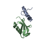


 PDBj
PDBj


 Assembly
 HNCA
HNCA Sample preparation
 Processing