Mass: 40.078 Da / Num. of mol.: 1 / Source method: obtained synthetically / Formula: Ca
-
Experimental details
-
Experiment
Experiment
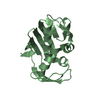
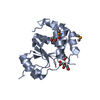
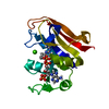
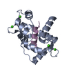
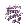
Method: SOLUTION NMR Details: EF-hand and SAM domain structural organization for calcium-loaded stromal interaction molecule 1
NMR experiment
Conditions-ID
Experiment-ID
Solution-ID
Type
1
1
1
2D 1H-15N HSQC
1
2
1
3DCBCA(CO)NH
1
3
1
3D HN(CA)CB
1
4
1
3D HNCO
1
5
1
3DH(CCO)NH
1
6
1
3DC(CO)NH
1
7
1
3D 1H-15N NOESY
2
8
2
3D (H)CCH-TOCSY
2
9
2
2D 1H-13C HSQC
2
10
2
3D 1H-13C NOESY
-
Sample preparation
Details
Solution-ID
Contents
Solvent system
1
0.5-1.0 mM [U-100% 13C; U-100% 15N] stromal interaction molecule 1, 20 mM TRIS, 100 mM sodium chloride, 5 mM CALCIUM ION, 95% H2O/5% D2O
95% H2O/5% D2O
2
0.5-1.0 mM [U-100% 13C; U-100% 15N] stromal interaction molecule 1, 20 mM [U-99% 2H] TRIS, 100 mM sodium chloride, 5 mM CALCIUM ION, 100% D2O
100% D2O
Sample
Conc. (mg/ml)
Component
Isotopic labeling
Solution-ID
0.5mM
stromalinteractionmolecule1
[U-100% 13C; U-100% 15N]
1
20mM
TRIS
1
100mM
sodiumchloride
1
5mM
CALCIUMION
1
0.5mM
stromalinteractionmolecule1
[U-100% 13C; U-100% 15N]
2
20mM
TRIS
[U-99% 2H]
2
100mM
sodiumchloride
2
5mM
CALCIUMION
2
Sample conditions
Conditions-ID
Ionic strength
pH
Pressure (kPa)
Temperature (K)
1
105
8
1atm
293K
2
105
1atm
293K
-
NMR measurement
NMR spectrometer
Type
Manufacturer
Model
Field strength (MHz)
Spectrometer-ID
Varian INOVA
Varian
INOVA
500
1
Bruker Avance
Bruker
AVANCE
800
2
-
Processing
NMR software
Name
Version
Developer
Classification
CNS
v1.1
BrungerA. T. et.al.
refinement
CYANA
v2.1
Guntert, MumenthalerandWuthrich
structuresolution
XEASY
Bartelsetal.
chemicalshiftassignment
NMRPipe
Delaglio, Grzesiek, Vuister, Zhu, PfeiferandBax
processing
Refinement
Method: simulated annealing, simulated annealing / Software ordinal: 1 Details: Water refinement and violation analysis was performed using the RECOORD scripts in CNS v1.1 (Nederveen et al, 2005).
NMR constraints
NOE constraints total: 3066 / NOE intraresidue total count: 796 / NOE long range total count: 802 / NOE medium range total count: 781 / NOE sequential total count: 687 / Hydrogen bond constraints total count: 106 / Protein phi angle constraints total count: 91 / Protein psi angle constraints total count: 91
NMR representative
Selection criteria: lowest energy
NMR ensemble
Conformer selection criteria: 20 structures for lowest energy Conformers calculated total number: 250 / Conformers submitted total number: 20
+
About Yorodumi
-
News
-
Feb 9, 2022. New format data for meta-information of EMDB entries
New format data for meta-information of EMDB entries
Version 3 of the EMDB header file is now the official format.
The previous official version 1.9 will be removed from the archive.
In the structure databanks used in Yorodumi, some data are registered as the other names, "COVID-19 virus" and "2019-nCoV". Here are the details of the virus and the list of structure data.
Jan 31, 2019. EMDB accession codes are about to change! (news from PDBe EMDB page)
EMDB accession codes are about to change! (news from PDBe EMDB page)
The allocation of 4 digits for EMDB accession codes will soon come to an end. Whilst these codes will remain in use, new EMDB accession codes will include an additional digit and will expand incrementally as the available range of codes is exhausted. The current 4-digit format prefixed with “EMD-” (i.e. EMD-XXXX) will advance to a 5-digit format (i.e. EMD-XXXXX), and so on. It is currently estimated that the 4-digit codes will be depleted around Spring 2019, at which point the 5-digit format will come into force.
The EM Navigator/Yorodumi systems omit the EMD- prefix.
Related info.:Q: What is EMD? / ID/Accession-code notation in Yorodumi/EM Navigator
Yorodumi is a browser for structure data from EMDB, PDB, SASBDB, etc.
This page is also the successor to EM Navigator detail page, and also detail information page/front-end page for Omokage search.
The word "yorodu" (or yorozu) is an old Japanese word meaning "ten thousand". "mi" (miru) is to see.
Related info.:EMDB / PDB / SASBDB / Comparison of 3 databanks / Yorodumi Search / Aug 31, 2016. New EM Navigator & Yorodumi / Yorodumi Papers / Jmol/JSmol / Function and homology information / Changes in new EM Navigator and Yorodumi
 Movie
Movie Controller


 Open data
 Basic information
 Components
 Keywords
 Function and homology information
 Homo sapiens (human)
 Authors
 Citation
 Structure visualization
 Downloads & links
 Other downloads
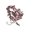


 PDBj
PDBj


 Assembly


 Mass: 40.078 Da / Num. of mol.: 1 / Source method: obtained synthetically / Formula: Ca
 HSQC
HSQC Sample preparation
 Processing