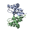
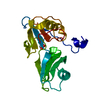
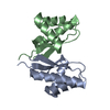
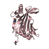
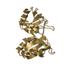
登録情報 データベース : PDB / ID : 2jziタイトル Structure of Calmodulin complexed with the Calmodulin Binding Domain of Calcineurin Calmodulin Serine/threonine-protein phosphatase 2B catalytic subunit alpha isoform キーワード / / / / / / / / / / / / / 機能・相同性 分子機能 ドメイン・相同性 構成要素
/ / / / / / / / / / / / / / / / / / / / / / / / / / / / / / / / / / / / / / / / / / / / / / / / / / / / / / / / / / / / / / / / / / / / / / / / / / / / / / / / / / / / / / / / / / / / / / / / / / / / / / / / / / / / / / / / / / / / / / / / / / / / / / / / / / / / / / / / / / 生物種 Homo sapiens (ヒト)手法 / データ登録者 Chyan, C. / Huang, J. / Irene, D. / Lin, T. ジャーナル : To be Published タイトル : Structure of Calmodulin complexed with the Calmodulin Binding Domain of Calcineurin著者 : Chyan, C. / Huang, J. / Irene, D. / Lin, T. 履歴 登録 2008年1月9日 登録サイト / 処理サイト 改定 1.0 2009年1月13日 Provider / タイプ 改定 1.1 2011年7月13日 Group 改定 1.2 2022年3月16日 Group / Database references / Derived calculationsカテゴリ database_2 / pdbx_nmr_spectrometer ... database_2 / pdbx_nmr_spectrometer / pdbx_struct_assembly / pdbx_struct_conn_angle / pdbx_struct_oper_list / struct_conn / struct_site Item _database_2.pdbx_DOI / _database_2.pdbx_database_accession ... _database_2.pdbx_DOI / _database_2.pdbx_database_accession / _pdbx_nmr_spectrometer.model / _pdbx_struct_conn_angle.ptnr1_auth_comp_id / _pdbx_struct_conn_angle.ptnr1_auth_seq_id / _pdbx_struct_conn_angle.ptnr1_label_atom_id / _pdbx_struct_conn_angle.ptnr1_label_comp_id / _pdbx_struct_conn_angle.ptnr1_label_seq_id / _pdbx_struct_conn_angle.ptnr3_auth_comp_id / _pdbx_struct_conn_angle.ptnr3_auth_seq_id / _pdbx_struct_conn_angle.ptnr3_label_atom_id / _pdbx_struct_conn_angle.ptnr3_label_comp_id / _pdbx_struct_conn_angle.ptnr3_label_seq_id / _pdbx_struct_conn_angle.value / _struct_conn.pdbx_dist_value / _struct_conn.ptnr1_auth_comp_id / _struct_conn.ptnr1_auth_seq_id / _struct_conn.ptnr1_label_atom_id / _struct_conn.ptnr1_label_comp_id / _struct_conn.ptnr1_label_seq_id / _struct_conn.ptnr2_auth_seq_id / _struct_conn.ptnr2_label_asym_id / _struct_site.pdbx_auth_asym_id / _struct_site.pdbx_auth_comp_id / _struct_site.pdbx_auth_seq_id 改定 1.3 2024年5月29日 Group / カテゴリ / chem_comp_bond
すべて表示 表示を減らす
 ムービー
ムービー コントローラー


 データを開く
 基本情報
基本情報 要素
要素 キーワード
キーワード 機能・相同性情報
 Homo sapiens (ヒト)
 データ登録者
 引用
引用 構造の表示
構造の表示 ダウンロードとリンク
 その他のダウンロード
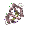


 PDBj
PDBj


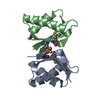
 集合体
集合体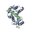


 分子量: 40.078 Da / 分子数: 4 / 由来タイプ: 合成 / 式: Ca
 HSQC
HSQC 試料調製
試料調製 解析
解析