 Movie
Movie Controller

[English] 日本語
 Yorodumi
- PDB-2jl4: Holo structure of Maleyl Pyruvate Isomerase, a bacterial glutathi... -
+ Open data

- Basic information
| Entry | Database: PDB / ID: 2jl4 | ||||||
|---|---|---|---|---|---|---|---|
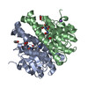
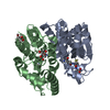
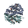
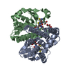
| Title | Holo structure of Maleyl Pyruvate Isomerase, a bacterial glutathione- s-transferase in Zeta class | ||||||
 Components Components | MALEYLPYRUVATE ISOMERASE | ||||||
 Keywords Keywords | ISOMERASE / GLUTATHIONE-S-TRANSFERASE / GST / PLASMID / PYRUVATE / BACTERIAL / BIODEGRADATION / MALEYL PYRUVATE / FUMARYL PYRUVATE | ||||||
| Function / homology |  Function and homology information Function and homology informationmaleylpyruvate isomerase / maleylpyruvate isomerase activity / naphthalene catabolic process / maleylacetoacetate isomerase activity / L-phenylalanine catabolic process / glutathione transferase activity / glutathione metabolic process / cytoplasm Similarity search - Function | ||||||
| Biological species |  RALSTONIA SP. U2 (bacteria) RALSTONIA SP. U2 (bacteria) | ||||||
| Method |  X-RAY DIFFRACTION / SYNCHROTRON / MOLECULAR REPLACEMENT / Resolution: 2.3 Å X-RAY DIFFRACTION / SYNCHROTRON / MOLECULAR REPLACEMENT / Resolution: 2.3 Å | ||||||
 Authors Authors | Shoemark, D.K. / Y Zhou, N. / Williams, P.A. / Hadfield, A.T. | ||||||
 Citation Citation | Journal: J.Mol.Biol. / Year: 2008 Title: Structure of Bacterial Glutathione-S-Transferase Maleyl Pyruvate Isomerase and Implications for Mechanism of Isomerisation. Authors: Marsh, M. / Shoemark, D.K. / Jacob, A. / Robinson, C. / Cahill, B. / Zhou, N.Y. / Williams, P.A. / Hadfield, A.T. #1: Journal: J.Bacteriol. / Year: 1998 Title: A Gene Cluster Encoding Steps in Conversion of Naphthalene to Gentisate in Pseudomonas Sp. Strain U2. Authors: Fuenmayor, S.L. / Wild, M. / Boyes, A.L. / Williams, P.A. | ||||||
| History |
|
- Structure visualization
| Structure viewer | Molecule: MolmilJmol/JSmol |
|---|
- Downloads & links
-Download
| PDBx/mmCIF format | 2jl4.cif.gz | 106.6 KB | Display | PDBx/mmCIF format |
|---|---|---|---|---|
| PDB format | pdb2jl4.ent.gz | 81.7 KB | Display | PDB format |
| PDBx/mmJSON format | 2jl4.json.gz | Tree view | PDBx/mmJSON format | |
| Others |  Other downloads Other downloads |
-Validation report
| Arichive directory | https://data.pdbj.org/pub/pdb/validation_reports/jl/2jl4ftp://data.pdbj.org/pub/pdb/validation_reports/jl/2jl4 | HTTPS FTP |
|---|
-Related structure data
| Related structure data |  2v6kC  1fw1S C: citing same article ( S: Starting model for refinement |
|---|---|
| Similar structure data |


-Links
 PDBj
PDBj

- Assembly
| Deposited unit | 
| ||||||||
|---|---|---|---|---|---|---|---|---|---|
| 1 |
| ||||||||
| Unit cell |
| ||||||||
| Noncrystallographic symmetry (NCS) | NCS oper: (Code: given Matrix: (-1, -0.0005, 0.00085), Vector: |
-Components
| #1: Protein | Mass: 23660.986 Da / Num. of mol.: 2 Source method: isolated from a genetically manipulated source Source: (gene. exp.) RALSTONIA SP. U2 (bacteria) / Description: VENEZUALAN OIL FIELDS / Production host: #2: Chemical |   Mass: 307.323 Da / Num. of mol.: 2 / Source method: obtained synthetically / Formula: C10H17N3O6S Mass: 307.323 Da / Num. of mol.: 2 / Source method: obtained synthetically / Formula: C10H17N3O6S#3: Water | ChemComp-HOH / |  Mass: 18.015 Da / Num. of mol.: 438 / Source method: isolated from a natural source / Formula: H2O Mass: 18.015 Da / Num. of mol.: 438 / Source method: isolated from a natural source / Formula: H2OSequence details | N-TERMINAL LYSINE VISIBLE WHICH COMES FROM HIS-TAG | |
|---|
-Experimental details
-Experiment
| Experiment | Method: X-RAY DIFFRACTION / Number of used crystals: 1 |
|---|
- Sample preparation
| Crystal | Density Matthews: 2.28 Å3/Da / Density % sol: 46 % / Description: NONE |
|---|---|
| Crystal grow | pH: 7.5 Details: 20% PEG 10K IN 0.1 M HEPES BUFFER AT PH 7.5, PROTEIN IN 20MM TRIS (PH 7.4), 2 MM GLUTATHIONE, 1 MM DTT AND 20 MM IMIDAZOLE |
-Data collection
| Diffraction | Mean temperature: 100 K |
|---|---|
| Diffraction source | Source: SYNCHROTRON / Site: SRS  / Beamline: PX14.2 / Wavelength: 1 / Beamline: PX14.2 / Wavelength: 1 |
| Detector | Type: ADSC CCD / Detector: CCD / Date: Feb 1, 2003 |
| Radiation | Protocol: SINGLE WAVELENGTH / Monochromatic (M) / Laue (L): M / Scattering type: x-ray |
| Radiation wavelength | Wavelength: 1 Å / Relative weight: 1 |
| Reflection | Resolution: 2.3→30 Å / Num. obs: 19625 / % possible obs: 97.3 % / Observed criterion σ(I): 0 / Redundancy: 4.3 % / Rmerge(I) obs: 0.12 |
| Reflection shell | Highest resolution: 2.3 Å / Rmerge(I) obs: 0.33 / % possible all: 93.8 |
- Processing
| Software |
| ||||||||||||||||||||||||||||||||||||||||||||||||||||||||||||||||||||||||||||||||||||||||||||||||||||||||||||||||||||||||||||||||||||||||||||||||||||||||||||||||||||||||||||||||||||||
|---|---|---|---|---|---|---|---|---|---|---|---|---|---|---|---|---|---|---|---|---|---|---|---|---|---|---|---|---|---|---|---|---|---|---|---|---|---|---|---|---|---|---|---|---|---|---|---|---|---|---|---|---|---|---|---|---|---|---|---|---|---|---|---|---|---|---|---|---|---|---|---|---|---|---|---|---|---|---|---|---|---|---|---|---|---|---|---|---|---|---|---|---|---|---|---|---|---|---|---|---|---|---|---|---|---|---|---|---|---|---|---|---|---|---|---|---|---|---|---|---|---|---|---|---|---|---|---|---|---|---|---|---|---|---|---|---|---|---|---|---|---|---|---|---|---|---|---|---|---|---|---|---|---|---|---|---|---|---|---|---|---|---|---|---|---|---|---|---|---|---|---|---|---|---|---|---|---|---|---|---|---|---|---|
| Refinement | Method to determine structure: MOLECULAR REPLACEMENT Starting model: PDB ENTRY 1FW1 Resolution: 2.3→27.63 Å / Cor.coef. Fo:Fc: 0.967 / Cor.coef. Fo:Fc free: 0.936 / SU B: 8.247 / SU ML: 0.204 / Cross valid method: THROUGHOUT / ESU R: 0.54 / ESU R Free: 0.286 / Stereochemistry target values: MAXIMUM LIKELIHOOD / Details: HYDROGENS HAVE BEEN ADDED IN THE RIDING POSITIONS.
| ||||||||||||||||||||||||||||||||||||||||||||||||||||||||||||||||||||||||||||||||||||||||||||||||||||||||||||||||||||||||||||||||||||||||||||||||||||||||||||||||||||||||||||||||||||||
| Solvent computation | Ion probe radii: 0.8 Å / Shrinkage radii: 0.8 Å / VDW probe radii: 1.4 Å / Solvent model: BABINET MODEL WITH MASK | ||||||||||||||||||||||||||||||||||||||||||||||||||||||||||||||||||||||||||||||||||||||||||||||||||||||||||||||||||||||||||||||||||||||||||||||||||||||||||||||||||||||||||||||||||||||
| Displacement parameters | Biso mean: 32.37 Å2
| ||||||||||||||||||||||||||||||||||||||||||||||||||||||||||||||||||||||||||||||||||||||||||||||||||||||||||||||||||||||||||||||||||||||||||||||||||||||||||||||||||||||||||||||||||||||
| Refinement step | Cycle: LAST / Resolution: 2.3→27.63 Å
| ||||||||||||||||||||||||||||||||||||||||||||||||||||||||||||||||||||||||||||||||||||||||||||||||||||||||||||||||||||||||||||||||||||||||||||||||||||||||||||||||||||||||||||||||||||||
| Refine LS restraints |
|