 Movie
Movie Controller

[English] 日本語
 Yorodumi
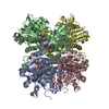
- PDB-2jjx: THE CRYSTAL STRUCTURE OF UMP KINASE FROM BACILLUS ANTHRACIS (BA1797) -
+ Open data

- Basic information
| Entry | Database: PDB / ID: 2jjx | ||||||
|---|---|---|---|---|---|---|---|
| Title | THE CRYSTAL STRUCTURE OF UMP KINASE FROM BACILLUS ANTHRACIS (BA1797) | ||||||
 Components Components | URIDYLATE KINASE | ||||||
 Keywords Keywords | TRANSFERASE / STRUCTURAL GENOMICS / PYRIMIDINE BIOSYNTHESIS / ATP-BINDING / URIDYLATE KINASE / NUCLEOTIDE-BINDING / OPPF / PYRH / KINASE / CYTOPLASM / OXFORD PROTEIN PRODUCTION FACILITY (OPPF) / STRUCTURAL PROTEOMICS IN EUROPE (SPINE) | ||||||
| Function / homology |  Function and homology information Function and homology informationUMP kinase / UMP kinase activity / 'de novo' CTP biosynthetic process / UDP biosynthetic process / ATP binding / cytoplasm Similarity search - Function | ||||||
| Biological species |  | ||||||
| Method |  X-RAY DIFFRACTION / SYNCHROTRON / MOLECULAR REPLACEMENT / Resolution: 2.82 Å X-RAY DIFFRACTION / SYNCHROTRON / MOLECULAR REPLACEMENT / Resolution: 2.82 Å | ||||||
 Authors Authors | Meier, C. / Carter, L.G. / Mancini, E.J. / Owens, R.J. / Stuart, D.I. / Esnouf, R.M. / Oxford Protein Production Facility (OPPF) / Structural Proteomics in Europe (SPINE) | ||||||
 Citation Citation | Journal: J.Mol.Biol. / Year: 2008 Title: The Crystal Structure of Ump Kinase from Bacillus Anthracis (Ba1797) Reveals an Allosteric Nucleotide-Binding Site. Authors: Meier, C. / Carter, L.G. / Sainsbury, S. / Mancini, E.J. / Owens, R.J. / Stuart, D.I. / Esnouf, R.M. | ||||||
| History |
|
- Structure visualization
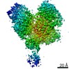
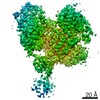
| Structure viewer | Molecule: MolmilJmol/JSmol |
|---|
- Downloads & links
-Download
| PDBx/mmCIF format | 2jjx.cif.gz | 159.8 KB | Display | PDBx/mmCIF format |
|---|---|---|---|---|
| PDB format | pdb2jjx.ent.gz | 126.4 KB | Display | PDB format |
| PDBx/mmJSON format | 2jjx.json.gz | Tree view | PDBx/mmJSON format | |
| Others |  Other downloads Other downloads |
-Validation report
| Arichive directory | https://data.pdbj.org/pub/pdb/validation_reports/jj/2jjxftp://data.pdbj.org/pub/pdb/validation_reports/jj/2jjx | HTTPS FTP |
|---|
-Related structure data
| Related structure data | 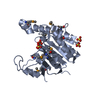 1z9dS S: Starting model for refinement |
|---|---|
| Similar structure data |


-Links
 PDBj
PDBj- Assembly
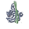
| Deposited unit | 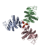
| ||||||||||||
|---|---|---|---|---|---|---|---|---|---|---|---|---|---|
| 1 | 
| ||||||||||||
| Unit cell |
| ||||||||||||
| Noncrystallographic symmetry (NCS) | NCS oper:
|
-Components
| #1: Protein | Mass: 28329.412 Da / Num. of mol.: 3 Source method: isolated from a genetically manipulated source Source: (gene. exp.) References: UniProt: Q81S73, UniProt: A0A6L7HKK4*PLUS, UMP kinase #2: Chemical |   Mass: 507.181 Da / Num. of mol.: 3 / Source method: obtained synthetically / Formula: C10H16N5O13P3 / Comment: ATP, energy-carrying molecule*YM Mass: 507.181 Da / Num. of mol.: 3 / Source method: obtained synthetically / Formula: C10H16N5O13P3 / Comment: ATP, energy-carrying molecule*YM#3: Chemical | ChemComp-MG / |   Mass: 24.305 Da / Num. of mol.: 1 / Source method: obtained synthetically / Formula: Mg Mass: 24.305 Da / Num. of mol.: 1 / Source method: obtained synthetically / Formula: Mg#4: Water | ChemComp-HOH / |  Mass: 18.015 Da / Num. of mol.: 142 / Source method: isolated from a natural source / Formula: H2O Mass: 18.015 Da / Num. of mol.: 142 / Source method: isolated from a natural source / Formula: H2O |
|---|
-Experimental details
-Experiment
| Experiment | Method: X-RAY DIFFRACTION / Number of used crystals: 1 |
|---|
- Sample preparation
| Crystal | Density Matthews: 2.49 Å3/Da / Density % sol: 50.6 % / Description: NONE |
|---|---|
| Crystal grow | pH: 8 Details: 0.2 M LITHIUM SULPHATE, 10% POLYETHYLENE GLYCOL 3000, 0.1 M IMIDAZOLE (PH 8.0) |
-Data collection
| Diffraction | Mean temperature: 100 K |
|---|---|
| Diffraction source | Source: SYNCHROTRON / Site: ESRF  / Beamline: BM14 / Wavelength: 0.886 / Beamline: BM14 / Wavelength: 0.886 |
| Detector | Type: MARRESEARCH / Detector: CCD / Date: Feb 27, 2005 |
| Radiation | Protocol: SINGLE WAVELENGTH / Monochromatic (M) / Laue (L): M / Scattering type: x-ray |
| Radiation wavelength | Wavelength: 0.886 Å / Relative weight: 1 |
| Reflection | Resolution: 2.82→50 Å / Num. obs: 21872 / % possible obs: 98.5 % / Observed criterion σ(I): 0 / Redundancy: 8.1 % / Rmerge(I) obs: 0.1 / Net I/σ(I): 12.6 |
| Reflection shell | Resolution: 2.82→2.93 Å / Redundancy: 7.1 % / Rmerge(I) obs: 0.77 / Mean I/σ(I) obs: 1.6 / % possible all: 98.8 |
- Processing
| Software |
| |||||||||||||||||||||||||||||||||||||||||||||||||||||||||
|---|---|---|---|---|---|---|---|---|---|---|---|---|---|---|---|---|---|---|---|---|---|---|---|---|---|---|---|---|---|---|---|---|---|---|---|---|---|---|---|---|---|---|---|---|---|---|---|---|---|---|---|---|---|---|---|---|---|---|
| Refinement | Method to determine structure: MOLECULAR REPLACEMENT Starting model: PDB ENTRY 1Z9D Resolution: 2.82→50 Å / Cross valid method: THROUGHOUT / σ(F): 0 Details: REGIONS 20-23 AND 171-176 IN EACH CHAIN ARE POORLY ORDERED AND CLASH BETWEEN SYMMETRY RELATED COPIES RESIDUE PHE106 IN A AND B CHAINS CLASH WITH EACH OTHER WHILE RESIDUE PHE106 IN C CHAIN ...Details: REGIONS 20-23 AND 171-176 IN EACH CHAIN ARE POORLY ORDERED AND CLASH BETWEEN SYMMETRY RELATED COPIES RESIDUE PHE106 IN A AND B CHAINS CLASH WITH EACH OTHER WHILE RESIDUE PHE106 IN C CHAIN HAS A SYMMETRY RELATED CLASH WITH ITSELF THE ACTIVE SITE OF EACH CHAIN CONTAINS UNMODELLED ELECTRON DENSITY PRESUMED TO BE ATP WEAKLY BOUND IN THE ABSENCE OF BOUND UMP.
| |||||||||||||||||||||||||||||||||||||||||||||||||||||||||
| Refinement step | Cycle: LAST / Resolution: 2.82→50 Å
| |||||||||||||||||||||||||||||||||||||||||||||||||||||||||
| Refine LS restraints |
|
