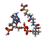
 Movie
Movie Controller

[English] 日本語
 Yorodumi
- PDB-2ji6: X-ray structure of Oxalyl-CoA decarboxylase in complex with 3-dea... -
+ Open data

- Basic information
| Entry | Database: PDB / ID: 2ji6 | ||||||
|---|---|---|---|---|---|---|---|
| Title | X-ray structure of Oxalyl-CoA decarboxylase in complex with 3-deaza- ThDP and oxalyl-CoA | ||||||
 Components Components | OXALYL-COA DECARBOXYLASE | ||||||
 Keywords Keywords | LYASE / FLAVOPROTEIN / DECARBOXYLASE / THIAMIN DIPHOSPHATE-DEPENDENT / SUBSTRATE COMPLEX / OXALATE DEGRADATION / THIAMINE PYROPHOSPHATE / NON- OXIDATIVE DECARBOXYLASE | ||||||
| Function / homology |  Function and homology information Function and homology informationoxalyl-CoA decarboxylase / oxalyl-CoA decarboxylase activity / oxalate catabolic process / fatty acid alpha-oxidation / thiamine pyrophosphate binding / ADP binding / magnesium ion binding / identical protein binding Similarity search - Function | ||||||
| Biological species |  OXALOBACTER FORMIGENES (bacteria) OXALOBACTER FORMIGENES (bacteria) | ||||||
| Method |  X-RAY DIFFRACTION / SYNCHROTRON / MOLECULAR REPLACEMENT / Resolution: 2.06 Å X-RAY DIFFRACTION / SYNCHROTRON / MOLECULAR REPLACEMENT / Resolution: 2.06 Å | ||||||
 Authors Authors | Berthold, C.L. / Toyota, C.G. / Moussatche, P. / Wood, M.D. / Leeper, F. / Richards, N.G.J. / Lindqvist, Y. | ||||||
 Citation Citation | Journal: Structure / Year: 2007 Title: Crystallographic Snapshots of Oxalyl-Coa Decarboxylase Give Insights Into Catalysis by Nonoxidative Thdp-Dependent Decarboxylases Authors: Berthold, C.L. / Toyota, C.G. / Moussatche, P. / Wood, M.D. / Leeper, F. / Richards, N.G.J. / Lindqvist, Y. | ||||||
| History |
|
- Structure visualization
| Structure viewer | Molecule: MolmilJmol/JSmol |
|---|
- Downloads & links
-Download
| PDBx/mmCIF format | 2ji6.cif.gz | 249.1 KB | Display | PDBx/mmCIF format |
|---|---|---|---|---|
| PDB format | pdb2ji6.ent.gz | 196.5 KB | Display | PDB format |
| PDBx/mmJSON format | 2ji6.json.gz | Tree view | PDBx/mmJSON format | |
| Others |  Other downloads Other downloads |
-Validation report
| Arichive directory | https://data.pdbj.org/pub/pdb/validation_reports/ji/2ji6ftp://data.pdbj.org/pub/pdb/validation_reports/ji/2ji6 | HTTPS FTP |
|---|
-Related structure data
| Related structure data |  2ji7C  2ji8C  2ji9C  2jibC  2c31S S: Starting model for refinement C: citing same article ( |
|---|---|
| Similar structure data |


-Links
 PDBj
PDBj


- Assembly
| Deposited unit | 
| ||||||||
|---|---|---|---|---|---|---|---|---|---|
| 1 | 
| ||||||||
| Unit cell |
| ||||||||
| Noncrystallographic symmetry (NCS) | NCS oper: (Code: given Matrix: (-1, 0.001919, -0.000965), Vector: |
-Components
-Protein , 1 types, 2 molecules AB
| #1: Protein | Mass: 60749.867 Da / Num. of mol.: 2 Source method: isolated from a genetically manipulated source Source: (gene. exp.) OXALOBACTER FORMIGENES (bacteria) / Plasmid: PET9A / Production host: |
|---|
-Non-polymers , 7 types, 920 molecules 


| #2: Chemical |  Mass: 423.318 Da / Num. of mol.: 2 / Source method: obtained synthetically / Formula: C13H19N3O7P2S Mass: 423.318 Da / Num. of mol.: 2 / Source method: obtained synthetically / Formula: C13H19N3O7P2S#3: Chemical |  Mass: 24.305 Da / Num. of mol.: 2 / Source method: obtained synthetically / Formula: Mg Mass: 24.305 Da / Num. of mol.: 2 / Source method: obtained synthetically / Formula: Mg#4: Chemical |  Mass: 427.201 Da / Num. of mol.: 2 / Source method: obtained synthetically / Formula: C10H15N5O10P2 / Comment: ADP, energy-carrying molecule*YM Mass: 427.201 Da / Num. of mol.: 2 / Source method: obtained synthetically / Formula: C10H15N5O10P2 / Comment: ADP, energy-carrying molecule*YM#5: Chemical |  Mass: 839.554 Da / Num. of mol.: 2 / Source method: obtained synthetically / Formula: C23H36N7O19P3S Mass: 839.554 Da / Num. of mol.: 2 / Source method: obtained synthetically / Formula: C23H36N7O19P3S#6: Chemical |  Mass: 150.173 Da / Num. of mol.: 2 / Source method: obtained synthetically / Formula: C6H14O4 Mass: 150.173 Da / Num. of mol.: 2 / Source method: obtained synthetically / Formula: C6H14O4#7: Chemical |  Mass: 282.334 Da / Num. of mol.: 2 / Source method: obtained synthetically / Formula: C11H26N2O6 / Comment: pH buffer*YM Mass: 282.334 Da / Num. of mol.: 2 / Source method: obtained synthetically / Formula: C11H26N2O6 / Comment: pH buffer*YM#8: Water | ChemComp-HOH / | Mass: 18.015 Da / Num. of mol.: 908 / Source method: isolated from a natural source / Formula: H2O |
|---|
-Experimental details
-Experiment
| Experiment | Method: X-RAY DIFFRACTION / Number of used crystals: 1 |
|---|
- Sample preparation
| Crystal | Density Matthews: 2.9 Å3/Da / Density % sol: 58 % / Description: NONE |
|---|
-Data collection
| Diffraction | Mean temperature: 100 K |
|---|---|
| Diffraction source | Source: SYNCHROTRON / Site: MAX II  / Beamline: I711 / Wavelength: 1.083 / Beamline: I711 / Wavelength: 1.083 |
| Detector | Type: MARRESEARCH / Detector: CCD / Date: Mar 19, 2006 / Details: VERTICALLY FOCUSING CYLINDRICAL MIRROR |
| Radiation | Monochromator: SINGLE ASYMMETRICALLY CUT SI(111) CRYSTAL WITH HORIZONTAL DIFFRACTION PLANE Protocol: SINGLE WAVELENGTH / Monochromatic (M) / Laue (L): M / Scattering type: x-ray |
| Radiation wavelength | Wavelength: 1.083 Å / Relative weight: 1 |
| Reflection | Resolution: 2.06→152 Å / Num. obs: 84850 / % possible obs: 96.2 % / Observed criterion σ(I): 0 / Redundancy: 6 % / Biso Wilson estimate: 24.88 Å2 / Rmerge(I) obs: 0.1 / Net I/σ(I): 11.8 |
| Reflection shell | Resolution: 2.06→2.17 Å / Redundancy: 4.4 % / Rmerge(I) obs: 0.5 / Mean I/σ(I) obs: 2.5 / % possible all: 74.2 |
- Processing
| Software |
| ||||||||||||||||||||||||||||||||||||||||||||||||||||||||||||||||||||||||||||||||||||||||||||||||||||||||||||||||||||||||||||||||||||||||||||||||||||||||||||||||||||||||||||||||||||||
|---|---|---|---|---|---|---|---|---|---|---|---|---|---|---|---|---|---|---|---|---|---|---|---|---|---|---|---|---|---|---|---|---|---|---|---|---|---|---|---|---|---|---|---|---|---|---|---|---|---|---|---|---|---|---|---|---|---|---|---|---|---|---|---|---|---|---|---|---|---|---|---|---|---|---|---|---|---|---|---|---|---|---|---|---|---|---|---|---|---|---|---|---|---|---|---|---|---|---|---|---|---|---|---|---|---|---|---|---|---|---|---|---|---|---|---|---|---|---|---|---|---|---|---|---|---|---|---|---|---|---|---|---|---|---|---|---|---|---|---|---|---|---|---|---|---|---|---|---|---|---|---|---|---|---|---|---|---|---|---|---|---|---|---|---|---|---|---|---|---|---|---|---|---|---|---|---|---|---|---|---|---|---|---|
| Refinement | Method to determine structure: MOLECULAR REPLACEMENT Starting model: PDB ENTRY 2C31 Resolution: 2.06→30 Å / Cor.coef. Fo:Fc: 0.961 / Cor.coef. Fo:Fc free: 0.942 / SU B: 8.892 / SU ML: 0.129 / TLS residual ADP flag: LIKELY RESIDUAL / Cross valid method: THROUGHOUT / ESU R: 0.176 / ESU R Free: 0.155 / Stereochemistry target values: MAXIMUM LIKELIHOOD / Details: HYDROGENS HAVE BEEN ADDED IN THE RIDING POSITIONS.
| ||||||||||||||||||||||||||||||||||||||||||||||||||||||||||||||||||||||||||||||||||||||||||||||||||||||||||||||||||||||||||||||||||||||||||||||||||||||||||||||||||||||||||||||||||||||
| Solvent computation | Ion probe radii: 0.8 Å / Shrinkage radii: 0.8 Å / VDW probe radii: 1.4 Å / Solvent model: MASK | ||||||||||||||||||||||||||||||||||||||||||||||||||||||||||||||||||||||||||||||||||||||||||||||||||||||||||||||||||||||||||||||||||||||||||||||||||||||||||||||||||||||||||||||||||||||
| Displacement parameters | Biso mean: 25.33 Å2
| ||||||||||||||||||||||||||||||||||||||||||||||||||||||||||||||||||||||||||||||||||||||||||||||||||||||||||||||||||||||||||||||||||||||||||||||||||||||||||||||||||||||||||||||||||||||
| Refinement step | Cycle: LAST / Resolution: 2.06→30 Å
| ||||||||||||||||||||||||||||||||||||||||||||||||||||||||||||||||||||||||||||||||||||||||||||||||||||||||||||||||||||||||||||||||||||||||||||||||||||||||||||||||||||||||||||||||||||||
| Refine LS restraints |
|