Monochromator: SI / Protocol: SINGLE WAVELENGTH / Monochromatic (M) / Laue (L): M / Scattering type: x-ray
Radiation wavelength
Wavelength: 0.9537 Å / Relative weight: 1
Reflection
Resolution: 3→25 Å / Num. obs: 14194 / % possible obs: 96.9 % / Observed criterion σ(I): 2.5 / Redundancy: 11 % / Rmerge(I) obs: 0.13 / Net I/σ(I): 19.5
Reflection shell
Resolution: 3→3.11 Å / Redundancy: 7.4 % / Rmerge(I) obs: 0.44 / Mean I/σ(I) obs: 2.5 / % possible all: 90.8
-
Processing
Software
Name
Version
Classification
REFMAC
5.2.0019
refinement
DENZO
datareduction
SCALEPACK
datascaling
Refinement
Method to determine structure: OTHER / Resolution: 3→25 Å / Cor.coef. Fo:Fc: 0.908 / Cor.coef. Fo:Fc free: 0.842 / SU B: 71.804 / SU ML: 0.589 / TLS residual ADP flag: LIKELY RESIDUAL / Cross valid method: THROUGHOUT / ESU R Free: 0.61 / Stereochemistry target values: MAXIMUM LIKELIHOOD / Details: HYDROGENS HAVE BEEN ADDED IN THE RIDING POSITIONS.
Rfactor
Num. reflection
% reflection
Selection details
Rfree
0.33
716
4.8 %
RANDOM
Rwork
0.248
-
-
-
obs
0.252
14194
96.8 %
-
Solvent computation
Ion probe radii: 0.8 Å / Shrinkage radii: 0.8 Å / VDW probe radii: 1.4 Å / Solvent model: MASK
 Movie
Movie Controller

 Yorodumi
 Open data
 Basic information
 Components
 Keywords
 Function and homology information
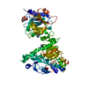
 BLUETONGUE VIRUS 10
 X-RAY DIFFRACTION /
  Authors
 Citation
 Structure visualization
 Downloads & links
 Other downloads


 PDBj
PDBj

 Assembly

 SPODOPTERA FRUGIPERDA (fall armyworm) / References: UniProt: P07132
 Mass: 523.180 Da / Num. of mol.: 1 / Source method: obtained synthetically / Formula: C10H16N5O14P3 / Comment: GTP, energy-carrying molecule*YM
 Mass: 151.126 Da / Num. of mol.: 2 / Source method: obtained synthetically / Formula: C5H5N5O
 Sample preparation
 / Beamline: BM14 / Wavelength: 0.9537
  Processing