| Entry | Database: PDB / ID: 6kms
|
|---|
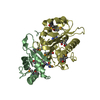
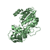
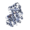
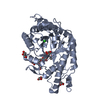
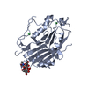
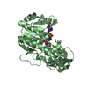
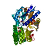
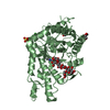
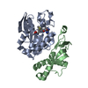
| Title | Crystal structure of human N6amt1-Trm112 in complex with SAM (space group I422) |
|---|
 Components Components | - Methyltransferase N6AMT1
- Multifunctional methyltransferase subunit TRM112-like protein
|
|---|
 Keywords Keywords | TRANSFERASE / methyltransferase / complex / protein translation / polypeptide release factor eRF1 |
|---|
| Function / homology |  Function and homology information Function and homology information
histone H4K12 methyltransferase activity / arsonoacetate metabolic process / protein-glutamine N-methyltransferase activity / arsenite methyltransferase activity / eRF1 methyltransferase complex / tRNA (m2G10) methyltransferase complex / tRNA methyltransferase activator activity / peptidyl-glutamine methylation / rRNA (guanine-N7)-methylation / toxin metabolic process ...histone H4K12 methyltransferase activity / arsonoacetate metabolic process / protein-glutamine N-methyltransferase activity / arsenite methyltransferase activity / eRF1 methyltransferase complex / tRNA (m2G10) methyltransferase complex / tRNA methyltransferase activator activity / peptidyl-glutamine methylation / rRNA (guanine-N7)-methylation / toxin metabolic process / site-specific DNA-methyltransferase (adenine-specific) activity / tRNA modification in the nucleus and cytosol / Methylation / protein methyltransferase activity / tRNA methylation / S-adenosylmethionine-dependent methyltransferase activity / S-adenosyl-L-methionine binding / positive regulation of rRNA processing / rRNA methylation / rRNA modification in the nucleus and cytosol / negative regulation of gene expression, epigenetic / Eukaryotic Translation Termination / maturation of LSU-rRNA / Transferases; Transferring one-carbon groups; Methyltransferases / transcription initiation-coupled chromatin remodeling / maturation of SSU-rRNA / positive regulation of cell growth / methylation / nucleic acid binding / protein heterodimerization activity / perinuclear region of cytoplasm / protein-containing complex / nucleoplasm / nucleus / cytosol / cytoplasmSimilarity search - Function Eukaryotic/archaeal PrmC-related / : / Multifunctional methyltransferase subunit Trm112 / Trm112-like / Trm112p-like protein / Methyltransferase small domain / Methyltransferase small domain / N-6 Adenine-specific DNA methylases signature. / DNA methylase, N-6 adenine-specific, conserved site / S-adenosyl-L-methionine-dependent methyltransferase superfamilySimilarity search - Domain/homology |
|---|
| Biological species |  Homo sapiens (human) Homo sapiens (human) |
|---|
| Method |  X-RAY DIFFRACTION / SYNCHROTRON / SAD / Resolution: 3.2 Å X-RAY DIFFRACTION / SYNCHROTRON / SAD / Resolution: 3.2 Å |
|---|
 Authors Authors | Li, W.J. / Shi, Y. / Zhang, T.L. / Ye, J. / Ding, J.P. |
|---|
| Funding support |  China, 2items China, 2items | Organization | Grant number | Country |
|---|
| National Natural Science Foundation of China | 31530013 | China | | National Natural Science Foundation of China | 31800622 | China |
|
|---|
 Citation Citation | Journal: Cell Discov / Year: 2019
Title: Structural insight into human N6amt1-Trm112 complex functioning as a protein methyltransferase.
Authors: Li, W. / Shi, Y. / Zhang, T. / Ye, J. / Ding, J. |
|---|
| History | | Deposition | Aug 1, 2019 | Deposition site: PDBJ / Processing site: PDBJ |
|---|
| Revision 1.0 | Sep 18, 2019 | Provider: repository / Type: Initial release |
|---|
| Revision 1.1 | Nov 6, 2019 | Group: Data collection / Database references / Category: citation / citation_author
Item: _citation.journal_volume / _citation.page_first ..._citation.journal_volume / _citation.page_first / _citation.page_last / _citation.pdbx_database_id_PubMed / _citation.title / _citation_author.identifier_ORCID / _citation_author.name |
|---|
| Revision 1.2 | Nov 20, 2024 | Group: Data collection / Database references ...Data collection / Database references / Refinement description / Structure summary
Category: chem_comp_atom / chem_comp_bond ...chem_comp_atom / chem_comp_bond / database_2 / pdbx_entry_details / pdbx_modification_feature / struct_ncs_dom_lim
Item: _database_2.pdbx_DOI / _database_2.pdbx_database_accession ..._database_2.pdbx_DOI / _database_2.pdbx_database_accession / _pdbx_entry_details.has_protein_modification / _struct_ncs_dom_lim.beg_auth_comp_id / _struct_ncs_dom_lim.beg_label_asym_id / _struct_ncs_dom_lim.beg_label_comp_id / _struct_ncs_dom_lim.beg_label_seq_id / _struct_ncs_dom_lim.end_auth_comp_id / _struct_ncs_dom_lim.end_label_asym_id / _struct_ncs_dom_lim.end_label_comp_id / _struct_ncs_dom_lim.end_label_seq_id |
|---|
|
|---|
 Movie
Movie Controller

 Yorodumi
 Open data
 Basic information
 Structure visualization
 Downloads & links
 Other downloads
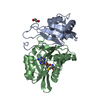


 PDBj
PDBj


 Assembly