Mass: 151.126 Da / Num. of mol.: 2 / Source method: obtained synthetically / Formula: C5H5N5O
Compound details
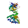
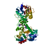
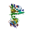
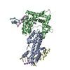
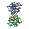
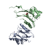
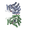
THE VP4 PROTEIN IS ONE OF THE FIVE PROTEINS (WITH VP1, VP3, VP6 AND VP7) WHICH FORMS THE INNER ...THE VP4 PROTEIN IS ONE OF THE FIVE PROTEINS (WITH VP1, VP3, VP6 AND VP7) WHICH FORMS THE INNER CAPSID OF THE VIRUS. VP4 CAPS VIRAL RNA.
-
Experimental details
-
Experiment
Experiment
Method: X-RAY DIFFRACTION / Number of used crystals: 1
-
Sample preparation
Crystal
Density Matthews: 2.26 Å3/Da / Density % sol: 45.68 % Description: THIS STRUCTURE IS A LIGAND (7MGDP) SOAK. THE ADOHCY (HIGH RESOLUTION) AND 7MGDP STRUCTURES WERE ISOMORPHOUS SO THIS STRUCTURE WAS SOLVED FROM THE ADOHCY - IT DID NOT REQUIR MOLECULAR ...Description: THIS STRUCTURE IS A LIGAND (7MGDP) SOAK. THE ADOHCY (HIGH RESOLUTION) AND 7MGDP STRUCTURES WERE ISOMORPHOUS SO THIS STRUCTURE WAS SOLVED FROM THE ADOHCY - IT DID NOT REQUIR MOLECULAR REPLACEMENT. VP4-7MGDP CRYSTALS ARE ISOMORPHOUS WITH VP4-ADOHCY CRYSTALS
Crystal grow
pH: 7.7 Details: 100 MM IMIDAZOLE PH 8.0, 2%-8% PROPAN-2-OL, 0-250 MM NACL, 50 MM DTT
In the structure databanks used in Yorodumi, some data are registered as the other names, "COVID-19 virus" and "2019-nCoV". Here are the details of the virus and the list of structure data.
Jan 31, 2019. EMDB accession codes are about to change! (news from PDBe EMDB page)
EMDB accession codes are about to change! (news from PDBe EMDB page)
The allocation of 4 digits for EMDB accession codes will soon come to an end. Whilst these codes will remain in use, new EMDB accession codes will include an additional digit and will expand incrementally as the available range of codes is exhausted. The current 4-digit format prefixed with “EMD-” (i.e. EMD-XXXX) will advance to a 5-digit format (i.e. EMD-XXXXX), and so on. It is currently estimated that the 4-digit codes will be depleted around Spring 2019, at which point the 5-digit format will come into force.
The EM Navigator/Yorodumi systems omit the EMD- prefix.
Related info.:Q: What is EMD? / ID/Accession-code notation in Yorodumi/EM Navigator
Yorodumi is a browser for structure data from EMDB, PDB, SASBDB, etc.
This page is also the successor to EM Navigator detail page, and also detail information page/front-end page for Omokage search.
The word "yorodu" (or yorozu) is an old Japanese word meaning "ten thousand". "mi" (miru) is to see.
Related info.:EMDB / PDB / SASBDB / Comparison of 3 databanks / Yorodumi Search / Aug 31, 2016. New EM Navigator & Yorodumi / Yorodumi Papers / Jmol/JSmol / Function and homology information / Changes in new EM Navigator and Yorodumi
 Movie
Movie Controller

 Yorodumi
 Open data
 Basic information
 Components
 Keywords
 Function and homology information
 BLUETONGUE VIRUS 10
 X-RAY DIFFRACTION /
  Authors
 Citation
 Structure visualization
 Downloads & links
 Other downloads


 PDBj
PDBj


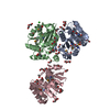
 Assembly
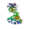

 SPODOPTERA FRUGIPERDA (fall armyworm) / References: UniProt: P07132, UniProt: Q65751*PLUS
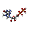
 Mass: 458.235 Da / Num. of mol.: 1 / Source method: obtained synthetically / Formula: C11H18N5O11P2
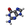
 Mass: 151.126 Da / Num. of mol.: 2 / Source method: obtained synthetically / Formula: C5H5N5O
 Sample preparation
 / Beamline: ID14-1 / Wavelength: 0.934
  Processing