| Entry | Database: PDB / ID: 2jbr
|
|---|
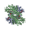
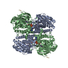
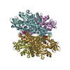
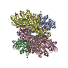
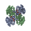
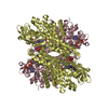
| Title | Structure of the monooxygenase component of p-hydroxyphenylacetate hydroxylase from Acinetobacter baumanni |
|---|
 Components Components | P-HYDROXYPHENYLACETATE HYDROXYLASE C2 OXYGENASE COMPONENT |
|---|
 Keywords Keywords | OXIDOREDUCTASE / FLAVOENZYME HYDROXYLASE |
|---|
| Function / homology |  Function and homology information Function and homology information
4-hydroxyphenylacetate 3-monooxygenase activity / 4-hydroxyphenylacetate 3-monooxygenase / fatty acid beta-oxidation using acyl-CoA dehydrogenase / acyl-CoA dehydrogenase activity / flavin adenine dinucleotide binding / cytoplasmSimilarity search - Function Acyl-CoA dehydrogenase, C-terminal domain / Acyl-CoA dehydrogenase, C-terminal domain / : / Butyryl-Coa Dehydrogenase, subunit A; domain 1 / Acyl-CoA dehydrogenase/oxidase, N-terminal domain / Butyryl-CoA Dehydrogenase, subunit A, domain 2 / Butyryl-CoA Dehydrogenase, subunit A; domain 2 / Acyl-CoA dehydrogenase/oxidase, N-terminal / Acyl-CoA dehydrogenase, N-terminal domain / Acyl-CoA dehydrogenase/oxidase, N-terminal domain superfamily ...Acyl-CoA dehydrogenase, C-terminal domain / Acyl-CoA dehydrogenase, C-terminal domain / : / Butyryl-Coa Dehydrogenase, subunit A; domain 1 / Acyl-CoA dehydrogenase/oxidase, N-terminal domain / Butyryl-CoA Dehydrogenase, subunit A, domain 2 / Butyryl-CoA Dehydrogenase, subunit A; domain 2 / Acyl-CoA dehydrogenase/oxidase, N-terminal / Acyl-CoA dehydrogenase, N-terminal domain / Acyl-CoA dehydrogenase/oxidase, N-terminal domain superfamily / Butyryl-CoA Dehydrogenase, subunit A, domain 3 / Acyl-CoA oxidase/dehydrogenase, middle domain superfamily / Acyl-CoA dehydrogenase/oxidase, N-terminal and middle domain superfamily / Acyl-CoA dehydrogenase-like, C-terminal / Butyryl-CoA Dehydrogenase, subunit A; domain 3 / Up-down Bundle / Beta Barrel / Orthogonal Bundle / Mainly Beta / Mainly AlphaSimilarity search - Domain/homology |
|---|
| Biological species |  ACINETOBACTER BAUMANNII (bacteria) ACINETOBACTER BAUMANNII (bacteria) |
|---|
| Method |  X-RAY DIFFRACTION / SYNCHROTRON / SAD / Resolution: 2.3 Å X-RAY DIFFRACTION / SYNCHROTRON / SAD / Resolution: 2.3 Å |
|---|
 Authors Authors | Alfieri, A. / Mattevi, A. |
|---|
 Citation Citation | Journal: Proc.Natl.Acad.Sci.USA / Year: 2007
Title: Structure of the Monooxygenase Component of a Two-Component Flavoprotein Monooxygenase.
Authors: Alfieri, A. / Fersini, F. / Ruangchan, N. / Prongjit, M. / Chaiyen, P. / Mattevi, A. |
|---|
| History | | Deposition | Dec 11, 2006 | Deposition site: PDBE / Processing site: PDBE |
|---|
| Revision 1.0 | Jan 23, 2007 | Provider: repository / Type: Initial release |
|---|
| Revision 1.1 | Jul 13, 2011 | Group: Advisory / Version format compliance |
|---|
| Revision 1.2 | May 8, 2024 | Group: Data collection / Database references / Other
Category: chem_comp_atom / chem_comp_bond ...chem_comp_atom / chem_comp_bond / database_2 / pdbx_database_status
Item: _database_2.pdbx_DOI / _database_2.pdbx_database_accession / _pdbx_database_status.status_code_sf |
|---|
|
|---|
 Movie
Movie Controller

 Yorodumi
 Open data
 Basic information
 Structure visualization
 Downloads & links
 Other downloads

 PDBj
PDBj Assembly
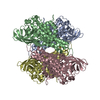
 Mass: 18.015 Da / Num. of mol.: 375 / Source method: isolated from a natural source / Formula: H2O
 Sample preparation
 / Beamline: ID14-1 / Wavelength: 1
  Processing