 Movie
Movie Controller

[English] 日本語
 Yorodumi
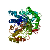
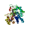
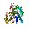
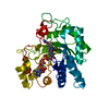
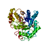
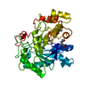
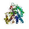
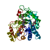
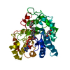
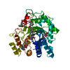
- PDB-2j8t: Human aldose reductase in complex with NADP and citrate at 0.82 a... -
+ Open data

- Basic information
| Entry | Database: PDB / ID: 2j8t | ||||||
|---|---|---|---|---|---|---|---|
| Title | Human aldose reductase in complex with NADP and citrate at 0.82 angstrom | ||||||
 Components Components | ALDO-KETO REDUCTASE FAMILY 1, MEMBER B1 | ||||||
 Keywords Keywords | OXIDOREDUCTASE / NADP / CITRATE | ||||||
| Function / homology |  Function and homology information Function and homology informationD-sorbitol metabolic process / glyceraldehyde oxidoreductase activity / Fructose biosynthesis / fructose biosynthetic process / L-glucuronate reductase activity / aldose reductase / D/L-glyceraldehyde reductase / glycerol dehydrogenase (NADP+) activity / C21-steroid hormone biosynthetic process / NADP-retinol dehydrogenase ...D-sorbitol metabolic process / glyceraldehyde oxidoreductase activity / Fructose biosynthesis / fructose biosynthetic process / L-glucuronate reductase activity / aldose reductase / D/L-glyceraldehyde reductase / glycerol dehydrogenase (NADP+) activity / C21-steroid hormone biosynthetic process / NADP-retinol dehydrogenase / Pregnenolone biosynthesis / allyl-alcohol dehydrogenase / allyl-alcohol dehydrogenase activity / Galactose catabolism / prostaglandin H2 endoperoxidase reductase activity / regulation of urine volume / all-trans-retinol dehydrogenase (NADP+) activity / metanephric collecting duct development / daunorubicin metabolic process / doxorubicin metabolic process / retinal dehydrogenase (NAD+) activity / epithelial cell maturation / aldose reductase (NADPH) activity / cellular hyperosmotic salinity response / renal water homeostasis / retinoid metabolic process / carbohydrate metabolic process / electron transfer activity / negative regulation of apoptotic process / mitochondrion / : / extracellular exosome / nucleoplasm / cytosol Similarity search - Function | ||||||
| Biological species |  HOMO SAPIENS (human) HOMO SAPIENS (human) | ||||||
| Method |  X-RAY DIFFRACTION / SYNCHROTRON / MOLECULAR REPLACEMENT / Resolution: 0.82 Å X-RAY DIFFRACTION / SYNCHROTRON / MOLECULAR REPLACEMENT / Resolution: 0.82 Å | ||||||
 Authors Authors | Biadene, M. / Hazemann, I. / Cousido, A. / Ginell, S. / Sheldrick, G.M. / Podjarny, A. / Schneider, T.R. | ||||||
 Citation Citation | Journal: Acta Crystallogr.,Sect.D / Year: 2007 Title: The Atomic Resolution Structure of Human Aldose Reductase Reveals that Rearrangement of a Bound Ligand Allows the Opening of the Safety-Belt Loop. Authors: Biadene, M. / Hazemann, I. / Cousido, A. / Ginell, S. / Joachimiak, A. / Sheldrick, G.M. / Podjarny, A. / Schneider, T.R. | ||||||
| History |
|
- Structure visualization
| Structure viewer | Molecule: MolmilJmol/JSmol |
|---|
- Downloads & links
-Download
| PDBx/mmCIF format | 2j8t.cif.gz | 183.3 KB | Display | PDBx/mmCIF format |
|---|---|---|---|---|
| PDB format | pdb2j8t.ent.gz | 144.2 KB | Display | PDB format |
| PDBx/mmJSON format | 2j8t.json.gz | Tree view | PDBx/mmJSON format | |
| Others |  Other downloads Other downloads |
-Validation report
| Arichive directory | https://data.pdbj.org/pub/pdb/validation_reports/j8/2j8tftp://data.pdbj.org/pub/pdb/validation_reports/j8/2j8t | HTTPS FTP |
|---|
-Related structure data
| Related structure data | 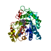 1el3S S: Starting model for refinement |
|---|---|
| Similar structure data |


-Links
 PDBj
PDBj


- Assembly
| Deposited unit | 
| ||||||||
|---|---|---|---|---|---|---|---|---|---|
| 1 |
| ||||||||
| Unit cell |
|
-Components
| #1: Protein | Mass: 35898.340 Da / Num. of mol.: 1 Source method: isolated from a genetically manipulated source Source: (gene. exp.) HOMO SAPIENS (human) / Production host:  References: UniProt: Q5U031, UniProt: P15121*PLUS, aldose reductase | ||
|---|---|---|---|
| #2: Chemical | ChemComp-NAP /   Mass: 743.405 Da / Num. of mol.: 1 / Source method: obtained synthetically / Formula: C21H28N7O17P3 Mass: 743.405 Da / Num. of mol.: 1 / Source method: obtained synthetically / Formula: C21H28N7O17P3 | ||
| #3: Chemical |   Mass: 189.100 Da / Num. of mol.: 2 / Source method: obtained synthetically / Formula: C6H5O7 Mass: 189.100 Da / Num. of mol.: 2 / Source method: obtained synthetically / Formula: C6H5O7#4: Water | ChemComp-HOH / |  Mass: 18.015 Da / Num. of mol.: 514 / Source method: isolated from a natural source / Formula: H2O Mass: 18.015 Da / Num. of mol.: 514 / Source method: isolated from a natural source / Formula: H2O |
-Experimental details
-Experiment
| Experiment | Method: X-RAY DIFFRACTION |
|---|
- Sample preparation
| Crystal | Density Matthews: 1.56 Å3/Da / Density % sol: 20.69 % |
|---|---|
| Crystal grow | pH: 5 Details: AS DESCRIBED IN LAMOUR ET AL. (1999) ACTA CRYSTALL. SECTION D 55: 721-723, pH 5.00 |
-Data collection
| Diffraction | Mean temperature: 10 K |
|---|---|
| Diffraction source | Source: SYNCHROTRON / Site: APS  / Beamline: 19-ID / Wavelength: 0.7999 / Beamline: 19-ID / Wavelength: 0.7999 |
| Detector | Type: ADSC CCD / Detector: CCD Details: 1.02-M FLAT MIRROR MADE OF ZERODUR PROVIDING VERTICAL FOCUSING AND REJECTION OF HARMONIC CONTAMINATION |
| Radiation | Monochromator: DOUBLE CRYSTAL MONOCHROMATOR UTILIZING A SI-111 AND SAGITAL HORIZONTAL FOCUSING Protocol: SINGLE WAVELENGTH / Monochromatic (M) / Laue (L): M / Scattering type: x-ray |
| Radiation wavelength | Wavelength: 0.7999 Å / Relative weight: 1 |
| Reflection | Resolution: 0.82→34.6 Å / Num. obs: 282677 / % possible obs: 94.4 % / Redundancy: 4.5 % / Rmerge(I) obs: 0.04 / Net I/σ(I): 26.1 |
| Reflection shell | Resolution: 0.82→0.85 Å / Redundancy: 3.3 % / Rmerge(I) obs: 0.08 / Mean I/σ(I) obs: 11.8 / % possible all: 90.4 |
- Processing
| Software |
| |||||||||||||||||||||||||||||||||
|---|---|---|---|---|---|---|---|---|---|---|---|---|---|---|---|---|---|---|---|---|---|---|---|---|---|---|---|---|---|---|---|---|---|---|
| Refinement | Method to determine structure: MOLECULAR REPLACEMENT Starting model: PDB ENTRY 1EL3 Resolution: 0.82→34.6 Å / Num. parameters: 32154 / Num. restraintsaints: 44811 / Cross valid method: FREE R-VALUE / σ(F): 0 / Stereochemistry target values: ENGH AND HUBER
| |||||||||||||||||||||||||||||||||
| Refine analyze | Num. disordered residues: 175 | |||||||||||||||||||||||||||||||||
| Refinement step | Cycle: LAST / Resolution: 0.82→34.6 Å
| |||||||||||||||||||||||||||||||||
| Refine LS restraints |
|
