 Movie
Movie Controller


+ Open data

- Basic information
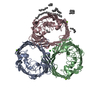
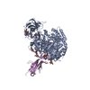
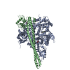
| Entry | Database: PDB / ID: 2j4u | ||||||
|---|---|---|---|---|---|---|---|
| Title | E.coli OmpC - camel Lactoferrin complex | ||||||
 Components Components |
| ||||||
 Keywords Keywords | MEMBRANE PROTEIN/HYDROLASE / MEMBRANE PROTEIN-HYDROLASE COMPLEX / IRON / OMPC / PORIN / COMPLEX / PROTEASE / HYDROLASE / MEMBRANE PROTEIN / ANTIACTERIAL PEPTIDE / ION TRANSPORT / IRON TRANSPORT / SERINE PROTEASE / TRANSPORT / LACTOFERRIN / GLYCOPROTEIN / METAL-BINDING | ||||||
| Function / homology |  Function and homology information Function and homology informationintermembrane phospholipid transfer / negative regulation of tumor necrosis factor (ligand) superfamily member 11 production / negative regulation of single-species biofilm formation in or on host organism / positive regulation of bone mineralization involved in bone maturation / negative regulation of osteoclast development / specific granule / negative regulation of lipopolysaccharide-mediated signaling pathway / positive regulation of chondrocyte proliferation / antifungal humoral response / regulation of tumor necrosis factor production ...intermembrane phospholipid transfer / negative regulation of tumor necrosis factor (ligand) superfamily member 11 production / negative regulation of single-species biofilm formation in or on host organism / positive regulation of bone mineralization involved in bone maturation / negative regulation of osteoclast development / specific granule / negative regulation of lipopolysaccharide-mediated signaling pathway / positive regulation of chondrocyte proliferation / antifungal humoral response / regulation of tumor necrosis factor production / bone morphogenesis / positive regulation of osteoblast proliferation / porin activity / pore complex / Hydrolases; Acting on peptide bonds (peptidases); Serine endopeptidases / positive regulation of osteoblast differentiation / regulation of cytokine production / ossification / innate immune response in mucosa / cell outer membrane / iron ion transport / recycling endosome / antibacterial humoral response / virus receptor activity / monoatomic ion transmembrane transport / early endosome / receptor-mediated virion attachment to host cell / iron ion binding / serine-type endopeptidase activity / DNA damage response / negative regulation of apoptotic process / proteolysis / : / metal ion binding / identical protein binding / plasma membrane Similarity search - Function | ||||||
| Biological species |   | ||||||
| Method |  X-RAY DIFFRACTION / SYNCHROTRON / MOLECULAR REPLACEMENT / Resolution: 2.99 Å X-RAY DIFFRACTION / SYNCHROTRON / MOLECULAR REPLACEMENT / Resolution: 2.99 Å | ||||||
 Authors Authors | Baalaji, S. / Acharya, R.K. / Singh, T.P. / Krishnaswamy, S. | ||||||
 Citation Citation | Journal: To be Published Title: Crystal Structure of the Membrane Protein Ompc Complex with Antibacterial Lactoferrin Authors: Baalaji, S. / Acharya, R.K. / Singh, T.P. / Krishnaswamy, S. | ||||||
| History |
| ||||||
| Remark 700 | SHEET DETERMINATION METHOD: DSSP THE SHEETS PRESENTED AS "QA" IN EACH CHAIN ON SHEET RECORDS BELOW ... SHEET DETERMINATION METHOD: DSSP THE SHEETS PRESENTED AS "QA" IN EACH CHAIN ON SHEET RECORDS BELOW IS ACTUALLY AN 17-STRANDED BARREL THIS IS REPRESENTED BY A 18-STRANDED SHEET IN WHICH THE FIRST AND LAST STRANDS ARE IDENTICAL. THE SHEETS PRESENTED AS "UA" IN EACH CHAIN ON SHEET RECORDS BELOW IS ACTUALLY AN 16-STRANDED BARREL THIS IS REPRESENTED BY A 17-STRANDED SHEET IN WHICH THE FIRST AND LAST STRANDS ARE IDENTICAL. THE SHEETS PRESENTED AS "VA" IN EACH CHAIN ON SHEET RECORDS BELOW IS ACTUALLY AN 16-STRANDED BARREL THIS IS REPRESENTED BY A 17-STRANDED SHEET IN WHICH THE FIRST AND LAST STRANDS ARE IDENTICAL. THE SHEETS PRESENTED AS "WA" IN EACH CHAIN ON SHEET RECORDS BELOW IS ACTUALLY AN 16-STRANDED BARREL THIS IS REPRESENTED BY A 17-STRANDED SHEET IN WHICH THE FIRST AND LAST STRANDS ARE IDENTICAL. THE SHEET STRUCTURE OF THIS MOLECULE IS BIFURCATED. IN ORDER TO REPRESENT THIS FEATURE IN THE SHEET RECORDS BELOW, TWO SHEETS ARE DEFINED. |
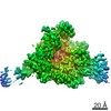
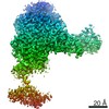
- Structure visualization
| Structure viewer | Molecule: MolmilJmol/JSmol |
|---|
- Downloads & links
-Download
| PDBx/mmCIF format | 2j4u.cif.gz | 416.6 KB | Display | PDBx/mmCIF format |
|---|---|---|---|---|
| PDB format | pdb2j4u.ent.gz | 331.9 KB | Display | PDB format |
| PDBx/mmJSON format | 2j4u.json.gz | Tree view | PDBx/mmJSON format | |
| Others |  Other downloads Other downloads |
-Validation report
| Arichive directory | https://data.pdbj.org/pub/pdb/validation_reports/j4/2j4uftp://data.pdbj.org/pub/pdb/validation_reports/j4/2j4u | HTTPS FTP |
|---|
-Related structure data


-Links
 PDBj
PDBj


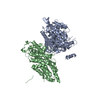
- Assembly
| Deposited unit | 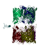
| |||||||||||||||||||||||||||||||||||||||||||||||||||||||||||||||||||||||||||||||||||||||||||||||||||||||||||||||||||||||||||||||||||||||||||
|---|---|---|---|---|---|---|---|---|---|---|---|---|---|---|---|---|---|---|---|---|---|---|---|---|---|---|---|---|---|---|---|---|---|---|---|---|---|---|---|---|---|---|---|---|---|---|---|---|---|---|---|---|---|---|---|---|---|---|---|---|---|---|---|---|---|---|---|---|---|---|---|---|---|---|---|---|---|---|---|---|---|---|---|---|---|---|---|---|---|---|---|---|---|---|---|---|---|---|---|---|---|---|---|---|---|---|---|---|---|---|---|---|---|---|---|---|---|---|---|---|---|---|---|---|---|---|---|---|---|---|---|---|---|---|---|---|---|---|---|---|
| 1 | 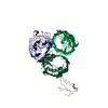
| |||||||||||||||||||||||||||||||||||||||||||||||||||||||||||||||||||||||||||||||||||||||||||||||||||||||||||||||||||||||||||||||||||||||||||
| 2 | 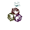
| |||||||||||||||||||||||||||||||||||||||||||||||||||||||||||||||||||||||||||||||||||||||||||||||||||||||||||||||||||||||||||||||||||||||||||
| Unit cell |
| |||||||||||||||||||||||||||||||||||||||||||||||||||||||||||||||||||||||||||||||||||||||||||||||||||||||||||||||||||||||||||||||||||||||||||
| Noncrystallographic symmetry (NCS) | NCS domain:
NCS domain segments: Component-ID: 1 / Beg auth comp-ID: ALA / Beg label comp-ID: ALA
NCS ensembles :
NCS oper:
|
-Components
| #1: Protein | Mass: 38336.242 Da / Num. of mol.: 6 / Fragment: RESIDUES 22-367 / Source method: isolated from a natural source / Details: LAB COLLECTION / Source: (natural) #2: Protein/peptide | Mass: 5203.188 Da / Num. of mol.: 2 / Fragment: N-TERM FRAGMENT, RESIDUES 20-64 / Source method: isolated from a natural source Details: ONLY 45 RESIDUES SEEN REMAINING COULD NOT BE LOCATED DUE TO DISORDER. Source: (natural) References: UniProt: Q9TUM0, Hydrolases; Acting on peptide bonds (peptidases); Serine endopeptidases Has protein modification | Y | Sequence details | SIGNAL PEPTIDE NOT PRESENT ONLY THE FIRST 45 RESIDUES ARE SEEN. THE REST COULD NOT BE LOCATED DUE TO DISORDER | |
|---|
-Experimental details
-Experiment
| Experiment | Method: X-RAY DIFFRACTION / Number of used crystals: 1 |
|---|
- Sample preparation
| Crystal | Density Matthews: 3.1 Å3/Da / Density % sol: 59.7 % |
|---|
-Data collection
| Diffraction | Mean temperature: 100 K |
|---|---|
| Diffraction source | Source: SYNCHROTRON / Site: SRS  / Beamline: PX14.2 / Wavelength: 0.9795 / Beamline: PX14.2 / Wavelength: 0.9795 |
| Detector | Type: ADSC QUANTUM 4 / Detector: CCD / Date: Aug 5, 2004 |
| Radiation | Protocol: SINGLE WAVELENGTH / Monochromatic (M) / Laue (L): M / Scattering type: x-ray |
| Radiation wavelength | Wavelength: 0.9795 Å / Relative weight: 1 |
| Reflection | Resolution: 3→50 Å / Num. obs: 55942 / % possible obs: 79 % / Observed criterion σ(I): 1 / Redundancy: 8.9 % / Rmerge(I) obs: 0.07 / Net I/σ(I): 12 |
| Reflection shell | Resolution: 3→3.11 Å / Redundancy: 7.6 % / Rmerge(I) obs: 0.25 / Mean I/σ(I) obs: 3.4 / % possible all: 73 |
- Processing
| Software |
| ||||||||||||||||||||||||||||||||||||||||||||||||||||||||||||||||||||||||||||||||||||||||||||||||||||||||||||||||||||||||||||||||||||||||||||||||||||||||||||||||||||||||||||||||||||||
|---|---|---|---|---|---|---|---|---|---|---|---|---|---|---|---|---|---|---|---|---|---|---|---|---|---|---|---|---|---|---|---|---|---|---|---|---|---|---|---|---|---|---|---|---|---|---|---|---|---|---|---|---|---|---|---|---|---|---|---|---|---|---|---|---|---|---|---|---|---|---|---|---|---|---|---|---|---|---|---|---|---|---|---|---|---|---|---|---|---|---|---|---|---|---|---|---|---|---|---|---|---|---|---|---|---|---|---|---|---|---|---|---|---|---|---|---|---|---|---|---|---|---|---|---|---|---|---|---|---|---|---|---|---|---|---|---|---|---|---|---|---|---|---|---|---|---|---|---|---|---|---|---|---|---|---|---|---|---|---|---|---|---|---|---|---|---|---|---|---|---|---|---|---|---|---|---|---|---|---|---|---|---|---|
| Refinement | Method to determine structure: MOLECULAR REPLACEMENT Starting model: PDB ENTRIES 1OSM, 1DTZ Resolution: 2.99→152.5 Å / Cor.coef. Fo:Fc: 0.916 / Cor.coef. Fo:Fc free: 0.857 / SU B: 15.655 / SU ML: 0.3 / Cross valid method: THROUGHOUT / ESU R Free: 0.518 / Stereochemistry target values: MAXIMUM LIKELIHOOD
| ||||||||||||||||||||||||||||||||||||||||||||||||||||||||||||||||||||||||||||||||||||||||||||||||||||||||||||||||||||||||||||||||||||||||||||||||||||||||||||||||||||||||||||||||||||||
| Solvent computation | Ion probe radii: 0.8 Å / Shrinkage radii: 0.8 Å / VDW probe radii: 1.2 Å / Solvent model: BABINET MODEL WITH MASK | ||||||||||||||||||||||||||||||||||||||||||||||||||||||||||||||||||||||||||||||||||||||||||||||||||||||||||||||||||||||||||||||||||||||||||||||||||||||||||||||||||||||||||||||||||||||
| Refinement step | Cycle: LAST / Resolution: 2.99→152.5 Å
| ||||||||||||||||||||||||||||||||||||||||||||||||||||||||||||||||||||||||||||||||||||||||||||||||||||||||||||||||||||||||||||||||||||||||||||||||||||||||||||||||||||||||||||||||||||||
| Refine LS restraints |
|