| Entry | Database: PDB / ID: 2j2i
|
|---|
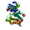
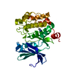
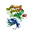
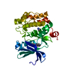
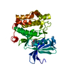
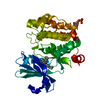
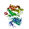
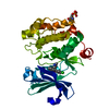
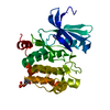
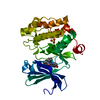
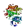
| Title | Crystal Structure of the humab PIM1 in complex with LY333531 |
|---|
 Components Components | PROTO-ONCOGENE SERINE/THREONINE-PROTEIN KINASE PIM-1 |
|---|
 Keywords Keywords | TRANSFERASE / NUCLEOTIDE-BINDING / ALTERNATIVE INITIATION / SERINE/THREONINE-PROTEIN KINASE / ATP-BINDING / METAL-BINDING / PROTO-ONCOGENE / KINASE / CANCER / LEUKEMIA / MANGANESE / NUCLEAR PROTEIN / PROTO- ONCOGENE / PHOSPHORYLATION |
|---|
| Function / homology |  Function and homology information Function and homology information
regulation of transmembrane transporter activity / positive regulation of cardioblast proliferation / positive regulation of cyclin-dependent protein serine/threonine kinase activity / regulation of hematopoietic stem cell proliferation / vitamin D receptor signaling pathway / cellular detoxification / STAT5 activation downstream of FLT3 ITD mutants / transcription factor binding / positive regulation of protein serine/threonine kinase activity / ribosomal small subunit binding ...regulation of transmembrane transporter activity / positive regulation of cardioblast proliferation / positive regulation of cyclin-dependent protein serine/threonine kinase activity / regulation of hematopoietic stem cell proliferation / vitamin D receptor signaling pathway / cellular detoxification / STAT5 activation downstream of FLT3 ITD mutants / transcription factor binding / positive regulation of protein serine/threonine kinase activity / ribosomal small subunit binding / : / positive regulation of cardiac muscle cell proliferation / positive regulation of brown fat cell differentiation / Signaling by FLT3 fusion proteins / positive regulation of TORC1 signaling / regulation of mitotic cell cycle / negative regulation of innate immune response / protein serine/threonine kinase activator activity / cellular response to type II interferon / protein autophosphorylation / manganese ion binding / Interleukin-4 and Interleukin-13 signaling / protein phosphorylation / non-specific serine/threonine protein kinase / protein stabilization / protein serine kinase activity / protein serine/threonine kinase activity / apoptotic process / negative regulation of apoptotic process / positive regulation of DNA-templated transcription / nucleolus / nucleoplasm / ATP binding / nucleus / plasma membrane / cytoplasm / cytosolSimilarity search - Function Serine/threonine-protein kinase pim-1/2/3 / : / Phosphorylase Kinase; domain 1 / Phosphorylase Kinase; domain 1 / Transferase(Phosphotransferase) domain 1 / Transferase(Phosphotransferase); domain 1 / Serine/threonine-protein kinase, active site / Serine/Threonine protein kinases active-site signature. / Protein kinase domain / Serine/Threonine protein kinases, catalytic domain ...Serine/threonine-protein kinase pim-1/2/3 / : / Phosphorylase Kinase; domain 1 / Phosphorylase Kinase; domain 1 / Transferase(Phosphotransferase) domain 1 / Transferase(Phosphotransferase); domain 1 / Serine/threonine-protein kinase, active site / Serine/Threonine protein kinases active-site signature. / Protein kinase domain / Serine/Threonine protein kinases, catalytic domain / Protein kinase, ATP binding site / Protein kinases ATP-binding region signature. / Protein kinase domain profile. / Protein kinase domain / Protein kinase-like domain superfamily / 2-Layer Sandwich / Orthogonal Bundle / Mainly Alpha / Alpha BetaSimilarity search - Domain/homology |
|---|
| Biological species |  HOMO SAPIENS (human) HOMO SAPIENS (human) |
|---|
| Method |  X-RAY DIFFRACTION / MOLECULAR REPLACEMENT / Resolution: 1.9 Å X-RAY DIFFRACTION / MOLECULAR REPLACEMENT / Resolution: 1.9 Å |
|---|
 Authors Authors | Debreczeni, J.E. / Bullock, A.N. / von Delft, F. / Sundstrom, M. / Arrowsmith, C. / Edwards, A. / Weigelt, J. / Knapp, S. |
|---|
 Citation Citation | Journal: Proc. Natl. Acad. Sci. U.S.A. / Year: 2007
Title: A systematic interaction map of validated kinase inhibitors with Ser/Thr kinases.
Authors: Fedorov, O. / Marsden, B. / Pogacic, V. / Rellos, P. / Muller, S. / Bullock, A.N. / Schwaller, J. / Sundstrom, M. / Knapp, S. |
|---|
| History | | Deposition | Aug 16, 2006 | Deposition site: PDBE / Processing site: PDBE |
|---|
| Revision 1.0 | Feb 13, 2007 | Provider: repository / Type: Initial release |
|---|
| Revision 1.1 | Jul 13, 2011 | Group: Advisory / Version format compliance |
|---|
| Revision 1.2 | Jan 24, 2018 | Group: Structure summary / Category: audit_author / Item: _audit_author.name |
|---|
| Revision 1.3 | Feb 28, 2018 | Group: Database references / Source and taxonomy / Category: citation / entity_src_gen
Item: _citation.journal_abbrev / _citation.journal_id_ISSN ..._citation.journal_abbrev / _citation.journal_id_ISSN / _citation.page_last / _citation.pdbx_database_id_DOI / _citation.title / _entity_src_gen.pdbx_host_org_cell_line / _entity_src_gen.pdbx_host_org_ncbi_taxonomy_id / _entity_src_gen.pdbx_host_org_scientific_name |
|---|
| Revision 1.4 | Apr 4, 2018 | Group: Data collection / Category: diffrn_source / Item: _diffrn_source.type |
|---|
| Revision 1.5 | Mar 6, 2019 | Group: Data collection / Experimental preparation / Category: exptl_crystal_grow / Item: _exptl_crystal_grow.temp |
|---|
| Revision 1.6 | May 8, 2019 | Group: Data collection / Experimental preparation / Category: exptl_crystal_grow / Item: _exptl_crystal_grow.method |
|---|
| Revision 1.7 | Dec 13, 2023 | Group: Data collection / Database references ...Data collection / Database references / Derived calculations / Other / Refinement description / Structure summary
Category: chem_comp / chem_comp_atom ...chem_comp / chem_comp_atom / chem_comp_bond / database_2 / entity / pdbx_database_status / pdbx_entity_nonpoly / pdbx_initial_refinement_model / struct_site
Item: _chem_comp.name / _database_2.pdbx_DOI ..._chem_comp.name / _database_2.pdbx_DOI / _database_2.pdbx_database_accession / _entity.pdbx_description / _pdbx_database_status.status_code_sf / _pdbx_entity_nonpoly.name / _struct_site.pdbx_auth_asym_id / _struct_site.pdbx_auth_comp_id / _struct_site.pdbx_auth_seq_id |
|---|
|
|---|
 Movie
Movie Controller


 Open data
 Basic information
 Structure visualization
 Downloads & links
 Other downloads


 PDBj
PDBj


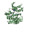
 Assembly


 Mass: 96.063 Da / Num. of mol.: 1 / Source method: obtained synthetically / Formula: SO4
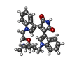
 Mass: 468.547 Da / Num. of mol.: 1 / Source method: obtained synthetically / Formula: C28H28N4O3
 Mass: 18.015 Da / Num. of mol.: 168 / Source method: isolated from a natural source / Formula: H2O
 Sample preparation
 Processing