TOXIN / BURKHOLDERIA PSEUDOMALLEI / BIPD / TTSS / T3SS / TYPE 3 SECRETION SYSTEM
Function / homology
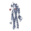
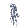
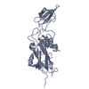
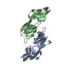
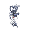
IpaD-like / IpaD-like / Type III secretion systems tip complex components / BipD-like superfamily / Type III secretion systems tip complex components / Up-down Bundle / extracellular region / Mainly Alpha / Translocator protein BipD
Function and homology information
Biological species
BURKHOLDERIA PSEUDOMALLEI (bacteria)
Method
X-RAY DIFFRACTION / SYNCHROTRON / MIRAS / Resolution: 2.6 Å
Journal: J Biol Chem / Year: 2007 Title: Self-chaperoning of the type III secretion system needle tip proteins IpaD and BipD. Authors: Steven Johnson / Pietro Roversi / Marianela Espina / Andrew Olive / Janet E Deane / Susan Birket / Terry Field / William D Picking / Ariel J Blocker / Edouard E Galyov / Wendy L Picking / Susan M Lea / Abstract: Bacteria expressing type III secretion systems (T3SS) have been responsible for the deaths of millions worldwide, acting as key virulence elements in diseases ranging from plague to typhoid fever. ...Bacteria expressing type III secretion systems (T3SS) have been responsible for the deaths of millions worldwide, acting as key virulence elements in diseases ranging from plague to typhoid fever. The T3SS is composed of a basal body, which traverses both bacterial membranes, and an external needle through which effector proteins are secreted. We report multiple crystal structures of two proteins that sit at the tip of the needle and are essential for virulence: IpaD from Shigella flexneri and BipD from Burkholderia pseudomallei. The structures reveal that the N-terminal domains of the molecules are intramolecular chaperones that prevent premature oligomerization, as well as sharing structural homology with proteins involved in eukaryotic actin rearrangement. Crystal packing has allowed us to construct a model for the tip complex that is supported by mutations designed using the structure.
#1: Journal: Acta Crystallogr.,Sect.F / Year: 2006 Title: Expression, Purification, Crystallization and Preliminary Crystallographic Analysis of Bipd, a Component of the Burkholderia Pseudomallei Type III Secretion System. Authors: Roversi, P. / Johnson, S. / Field, T. / Deane, J.E. / Galyov, E.E. / Lea, S.M.
Mass: 18.015 Da / Num. of mol.: 50 / Source method: isolated from a natural source / Formula: H2O
Sequence details
THE GS INITIAL RESIDUES COME FROM THE TAG, THE SEQUENCE STARTS AT BIPD RESIDUE 10
-
Experimental details
-
Experiment
Experiment
Method: X-RAY DIFFRACTION / Number of used crystals: 1
-
Sample preparation
Crystal
Density Matthews: 2.3 Å3/Da / Density % sol: 47 % Description: TWO PT ATOMS WERE FOUND IN K2PTCL4 ANOMALOUS DIFFERENCE PATTERSON MAPS USING SHELXD. THESE PT SITES ALLOWED LOCATION OF THE SE ATOMS IN A SEMET DERIVATIVE ANOMALOUS FOURIER DIFFERENCE MAP USING SHARP.
Crystal grow
pH: 8.5 / Details: 1 M TRISODIUM CITRATE, 10 MM SODIUM BORATE PH 8.5
In the structure databanks used in Yorodumi, some data are registered as the other names, "COVID-19 virus" and "2019-nCoV". Here are the details of the virus and the list of structure data.
Jan 31, 2019. EMDB accession codes are about to change! (news from PDBe EMDB page)
EMDB accession codes are about to change! (news from PDBe EMDB page)
The allocation of 4 digits for EMDB accession codes will soon come to an end. Whilst these codes will remain in use, new EMDB accession codes will include an additional digit and will expand incrementally as the available range of codes is exhausted. The current 4-digit format prefixed with “EMD-” (i.e. EMD-XXXX) will advance to a 5-digit format (i.e. EMD-XXXXX), and so on. It is currently estimated that the 4-digit codes will be depleted around Spring 2019, at which point the 5-digit format will come into force.
The EM Navigator/Yorodumi systems omit the EMD- prefix.
Related info.:Q: What is EMD? / ID/Accession-code notation in Yorodumi/EM Navigator
Yorodumi is a browser for structure data from EMDB, PDB, SASBDB, etc.
This page is also the successor to EM Navigator detail page, and also detail information page/front-end page for Omokage search.
The word "yorodu" (or yorozu) is an old Japanese word meaning "ten thousand". "mi" (miru) is to see.
Related info.:EMDB / PDB / SASBDB / Comparison of 3 databanks / Yorodumi Search / Aug 31, 2016. New EM Navigator & Yorodumi / Yorodumi Papers / Jmol/JSmol / Function and homology information / Changes in new EM Navigator and Yorodumi
 Movie
Movie Controller


 Open data
 Basic information
 Components
 Keywords
 Function and homology information
 BURKHOLDERIA PSEUDOMALLEI (bacteria)
 X-RAY DIFFRACTION /
  Authors
 Citation
 Structure visualization
 Downloads & links
 Other downloads
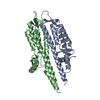


 PDBj
PDBj Assembly
 Mass: 18.015 Da / Num. of mol.: 50 / Source method: isolated from a natural source / Formula: H2O
 Sample preparation
 / Beamline: ID29 / Wavelength: 0.9778
  Processing