 Movie
Movie Controller


+ Open data

- Basic information
| Entry | Database: PDB / ID: 2ive | ||||||
|---|---|---|---|---|---|---|---|
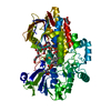
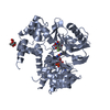
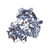
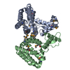
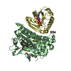
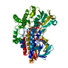
| Title | Structure of protoporphyrinogen oxidase from Myxococcus xanthus | ||||||
 Components Components | PROTOPORPHYRINOGEN OXIDASE | ||||||
 Keywords Keywords | OXIDOREDUCTASE / PROTOPORPHYRINOGEN OXIDASE / PORPHYRIN BIOSYNTHESIS / CHLOROPHYLL BIOSYNTHESIS / FAD / PORPHYRIA / FLAVOPROTEIN / HEME BIOSYNTHESIS / HAEM BIOSYNTHESIS | ||||||
| Function / homology |  Function and homology information Function and homology informationprotoporphyrinogen oxidase / protoporphyrinogen oxidase activity, oxygen as acceptor / : / plasma membrane / cytoplasm Similarity search - Function | ||||||
| Biological species |  MYXOCOCCUS XANTHUS (bacteria) MYXOCOCCUS XANTHUS (bacteria) | ||||||
| Method |  X-RAY DIFFRACTION / SYNCHROTRON / MOLECULAR REPLACEMENT / Resolution: 2.7 Å X-RAY DIFFRACTION / SYNCHROTRON / MOLECULAR REPLACEMENT / Resolution: 2.7 Å | ||||||
 Authors Authors | Corradi, H.R. / Corrigall, A.V. / Boix, E. / Mohan, C.G. / Sturrock, E.D. / Meissner, P.N. / Acharya, K.R. | ||||||
 Citation Citation | Journal: J.Biol.Chem. / Year: 2006 Title: Crystal Structure of Protoporphyrinogen Oxidase from Myxococcus Xanthus and its Complex with the Inhibitor Acifluorfen. Authors: Corradi, H.R. / Corrigall, A.V. / Boix, E. / Mohan, C.G. / Sturrock, E.D. / Meissner, P.N. / Acharya, K.R. | ||||||
| History |
|
- Structure visualization
| Structure viewer | Molecule: MolmilJmol/JSmol |
|---|
- Downloads & links
-Download
| PDBx/mmCIF format | 2ive.cif.gz | 185.3 KB | Display | PDBx/mmCIF format |
|---|---|---|---|---|
| PDB format | pdb2ive.ent.gz | 145.7 KB | Display | PDB format |
| PDBx/mmJSON format | 2ive.json.gz | Tree view | PDBx/mmJSON format | |
| Others |  Other downloads Other downloads |
-Validation report
| Arichive directory | https://data.pdbj.org/pub/pdb/validation_reports/iv/2iveftp://data.pdbj.org/pub/pdb/validation_reports/iv/2ive | HTTPS FTP |
|---|
-Related structure data
| Related structure data |  2ivdC  1sezS C: citing same article ( S: Starting model for refinement |
|---|---|
| Similar structure data |


-Links
 PDBj
PDBj
- Assembly
| Deposited unit | 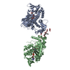
| ||||||||
|---|---|---|---|---|---|---|---|---|---|
| 1 | 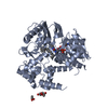
| ||||||||
| 2 | 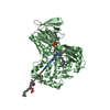
| ||||||||
| Unit cell |
| ||||||||
| Components on special symmetry positions |
| ||||||||
| Noncrystallographic symmetry (NCS) | NCS oper: (Code: given Matrix: (-0.68436, 0.72908, 0.01024), Vector: |
-Components
| #1: Protein | Mass: 50396.566 Da / Num. of mol.: 2 Source method: isolated from a genetically manipulated source Source: (gene. exp.) MYXOCOCCUS XANTHUS (bacteria) / Production host: #2: Chemical |   Mass: 785.550 Da / Num. of mol.: 2 / Source method: obtained synthetically / Formula: C27H33N9O15P2 / Comment: FAD*YM Mass: 785.550 Da / Num. of mol.: 2 / Source method: obtained synthetically / Formula: C27H33N9O15P2 / Comment: FAD*YM#3: Chemical | ChemComp-GOL /   Mass: 92.094 Da / Num. of mol.: 6 / Source method: obtained synthetically / Formula: C3H8O3 Mass: 92.094 Da / Num. of mol.: 6 / Source method: obtained synthetically / Formula: C3H8O3#4: Chemical |   Mass: 354.567 Da / Num. of mol.: 3 / Source method: obtained synthetically / Formula: C22H42O3 Mass: 354.567 Da / Num. of mol.: 3 / Source method: obtained synthetically / Formula: C22H42O3#5: Water | ChemComp-HOH / |  Mass: 18.015 Da / Num. of mol.: 146 / Source method: isolated from a natural source / Formula: H2O Mass: 18.015 Da / Num. of mol.: 146 / Source method: isolated from a natural source / Formula: H2OCompound details | PROTOPORPH | Sequence details | THE FIRST MET IN THE PPOX SEQUENCE WAS REPLACED BY THE HIS TAG IN THE CONSTRUCT, BUT THIS RESIDUE ...THE FIRST MET IN THE PPOX SEQUENCE WAS REPLACED BY THE HIS TAG IN THE CONSTRUCT, BUT THIS RESIDUE WAS NOT OBSERVED IN THE STRUCTURE | |
|---|
-Experimental details
-Experiment
| Experiment | Method: X-RAY DIFFRACTION / Number of used crystals: 1 |
|---|
- Sample preparation
| Crystal | Density Matthews: 3.74 Å3/Da / Density % sol: 66.85 % |
|---|---|
| Crystal grow | pH: 7.5 Details: 0.1M TRIS/HCL PH 7.5, 1.5M AMMONIUM SULPHATE, 20% GLYCEROL, 1% PEG 4000 |
-Data collection
| Diffraction | Mean temperature: 100 K |
|---|---|
| Diffraction source | Source: SYNCHROTRON / Site: SRS  / Beamline: PX14.2 / Wavelength: 0.978 / Beamline: PX14.2 / Wavelength: 0.978 |
| Detector | Type: ADSC CCD / Detector: CCD |
| Radiation | Protocol: SINGLE WAVELENGTH / Monochromatic (M) / Laue (L): M / Scattering type: x-ray |
| Radiation wavelength | Wavelength: 0.978 Å / Relative weight: 1 |
| Reflection | Resolution: 2.7→74.3 Å / Num. obs: 41382 / % possible obs: 100 % / Observed criterion σ(I): 2 / Redundancy: 7.5 % / Rmerge(I) obs: 0.14 / Net I/σ(I): 14.7 |
| Reflection shell | Resolution: 2.7→2.85 Å / Rmerge(I) obs: 0.57 / Mean I/σ(I) obs: 2.6 / % possible all: 99.8 |
- Processing
| Software |
| ||||||||||||||||||||||||||||||||||||||||||||||||||||||||||||||||||||||||||||||||||||||||||||||||||||||||||||||||||||||||||||||||||||||||||||||||||||||||||||||||||||||||||||||||||||||
|---|---|---|---|---|---|---|---|---|---|---|---|---|---|---|---|---|---|---|---|---|---|---|---|---|---|---|---|---|---|---|---|---|---|---|---|---|---|---|---|---|---|---|---|---|---|---|---|---|---|---|---|---|---|---|---|---|---|---|---|---|---|---|---|---|---|---|---|---|---|---|---|---|---|---|---|---|---|---|---|---|---|---|---|---|---|---|---|---|---|---|---|---|---|---|---|---|---|---|---|---|---|---|---|---|---|---|---|---|---|---|---|---|---|---|---|---|---|---|---|---|---|---|---|---|---|---|---|---|---|---|---|---|---|---|---|---|---|---|---|---|---|---|---|---|---|---|---|---|---|---|---|---|---|---|---|---|---|---|---|---|---|---|---|---|---|---|---|---|---|---|---|---|---|---|---|---|---|---|---|---|---|---|---|
| Refinement | Method to determine structure: MOLECULAR REPLACEMENT Starting model: PDB ENTRY 1SEZ Resolution: 2.7→74.33 Å / Cor.coef. Fo:Fc: 0.889 / Cor.coef. Fo:Fc free: 0.849 / SU B: 12.894 / SU ML: 0.262 / Cross valid method: THROUGHOUT / ESU R: 0.496 / ESU R Free: 0.323 / Stereochemistry target values: MAXIMUM LIKELIHOOD Details: HYDROGENS HAVE BEEN ADDED IN THE RIDING POSITIONS. RESIDUES 87-93 IN BOTH MOLECULES COULD NOT BE MODELLED VERY ACCURATELY DUE TO LOTS OF NOISE IN THE MAPS
| ||||||||||||||||||||||||||||||||||||||||||||||||||||||||||||||||||||||||||||||||||||||||||||||||||||||||||||||||||||||||||||||||||||||||||||||||||||||||||||||||||||||||||||||||||||||
| Solvent computation | Ion probe radii: 0.8 Å / Shrinkage radii: 0.8 Å / VDW probe radii: 1.2 Å / Solvent model: MASK | ||||||||||||||||||||||||||||||||||||||||||||||||||||||||||||||||||||||||||||||||||||||||||||||||||||||||||||||||||||||||||||||||||||||||||||||||||||||||||||||||||||||||||||||||||||||
| Displacement parameters | Biso mean: 30.6 Å2
| ||||||||||||||||||||||||||||||||||||||||||||||||||||||||||||||||||||||||||||||||||||||||||||||||||||||||||||||||||||||||||||||||||||||||||||||||||||||||||||||||||||||||||||||||||||||
| Refinement step | Cycle: LAST / Resolution: 2.7→74.33 Å
| ||||||||||||||||||||||||||||||||||||||||||||||||||||||||||||||||||||||||||||||||||||||||||||||||||||||||||||||||||||||||||||||||||||||||||||||||||||||||||||||||||||||||||||||||||||||
| Refine LS restraints |
|