[English] 日本語
Yorodumi
- PDB-2isy: Crystal structure of the nickel-activated two-domain iron-depende... -

+
Open data


ID or keywords:

Loading...

-
Basic information

Entry
Database: PDB / ID: 2isy
Title
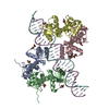
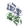
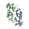
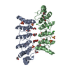
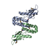
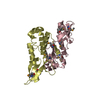
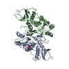
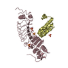
Crystal structure of the nickel-activated two-domain iron-dependent regulator (IdeR)
ComponentsIron-dependent repressor ideR
KeywordsTRANSCRIPTION / DNA-binding protein
Function / homology
Function and homology information


catechol-containing siderophore biosynthetic process / cobalt ion binding / cadmium ion binding / nickel cation binding / transition metal ion binding / peptidoglycan-based cell wall / ferrous iron binding / manganese ion binding / response to oxidative stress / protein dimerization activity ...catechol-containing siderophore biosynthetic process / cobalt ion binding / cadmium ion binding / nickel cation binding / transition metal ion binding / peptidoglycan-based cell wall / ferrous iron binding / manganese ion binding / response to oxidative stress / protein dimerization activity / iron ion binding / DNA-binding transcription factor activity / negative regulation of DNA-templated transcription / regulation of DNA-templated transcription / DNA binding / zinc ion binding / plasma membrane / cytoplasm
Similarity search - Function
Diphteria toxin repressor, SH3 domain / Diphteria toxin repressor SH3 domain / FeoA domain / Ferrous iron transporter, core domain / Ferrous iron transporter FeoA domain / FeoA / DtxR-type HTH domain profile. / DTXR-type HTH domain / Iron dependent repressor, N-terminal DNA binding domain / Iron dependent repressor, metal binding and dimerisation domain ...Diphteria toxin repressor, SH3 domain / Diphteria toxin repressor SH3 domain / FeoA domain / Ferrous iron transporter, core domain / Ferrous iron transporter FeoA domain / FeoA / DtxR-type HTH domain profile. / DTXR-type HTH domain / Iron dependent repressor, N-terminal DNA binding domain / Iron dependent repressor, metal binding and dimerisation domain / Iron dependent repressor / Iron dependent repressor, metal binding and dimerisation domain superfamily / Iron dependent repressor, metal binding and dimerisation domain / Helix-turn-helix diphteria tox regulatory element / Transcriptional repressor, C-terminal / Winged helix-like DNA-binding domain superfamily/Winged helix DNA-binding domain / Arc Repressor Mutant, subunit A / Winged helix DNA-binding domain superfamily / Winged helix-like DNA-binding domain superfamily / Orthogonal Bundle / Mainly Alpha
Similarity search - Domain/homology
NICKEL (II) ION / PHOSPHATE ION / Iron-dependent repressor IdeR / Iron-dependent repressor IdeR
Similarity search - Component
Biological speciesMycobacterium tuberculosis (bacteria)
MethodX-RAY DIFFRACTION / SYNCHROTRON / MOLECULAR REPLACEMENT / Resolution: 1.955 Å
AuthorsWisedchaisri, G. / Chou, C.J. / Wu, M. / Roach, C. / Rice, A.E. / Holmes, R.K. / Beeson, C. / Hol, W.G.
CitationJournal: Biochemistry / Year: 2007
Title: Crystal structures, metal activation, and DNA-binding properties of two-domain IdeR from Mycobacterium tuberculosis
Authors: Wisedchaisri, G. / Chou, C.J. / Wu, M. / Roach, C. / Rice, A.E. / Holmes, R.K. / Beeson, C. / Hol, W.G.
History
DepositionOct 18, 2006Deposition site: RCSB / Processing site: RCSB
Revision 1.0Feb 13, 2007Provider: repository / Type: Initial release
Revision 1.1May 1, 2008Group: Version format compliance
Revision 1.2Jul 13, 2011Group: Version format compliance
Revision 1.3Oct 18, 2017Group: Refinement description / Category: software
Revision 1.4Aug 30, 2023Group: Data collection / Database references ...Data collection / Database references / Derived calculations / Refinement description
Category: chem_comp_atom / chem_comp_bond ...chem_comp_atom / chem_comp_bond / database_2 / pdbx_initial_refinement_model / pdbx_struct_conn_angle / struct_conn / struct_ref_seq_dif / struct_site
Item: _database_2.pdbx_DOI / _database_2.pdbx_database_accession ..._database_2.pdbx_DOI / _database_2.pdbx_database_accession / _pdbx_struct_conn_angle.ptnr1_auth_comp_id / _pdbx_struct_conn_angle.ptnr1_auth_seq_id / _pdbx_struct_conn_angle.ptnr1_label_alt_id / _pdbx_struct_conn_angle.ptnr1_label_asym_id / _pdbx_struct_conn_angle.ptnr1_label_atom_id / _pdbx_struct_conn_angle.ptnr1_label_comp_id / _pdbx_struct_conn_angle.ptnr1_label_seq_id / _pdbx_struct_conn_angle.ptnr2_auth_seq_id / _pdbx_struct_conn_angle.ptnr2_label_asym_id / _pdbx_struct_conn_angle.ptnr3_auth_comp_id / _pdbx_struct_conn_angle.ptnr3_auth_seq_id / _pdbx_struct_conn_angle.ptnr3_label_alt_id / _pdbx_struct_conn_angle.ptnr3_label_asym_id / _pdbx_struct_conn_angle.ptnr3_label_atom_id / _pdbx_struct_conn_angle.ptnr3_label_comp_id / _pdbx_struct_conn_angle.ptnr3_label_seq_id / _pdbx_struct_conn_angle.value / _struct_conn.conn_type_id / _struct_conn.id / _struct_conn.pdbx_dist_value / _struct_conn.pdbx_leaving_atom_flag / _struct_conn.pdbx_ptnr1_label_alt_id / _struct_conn.pdbx_ptnr2_label_alt_id / _struct_conn.ptnr1_auth_asym_id / _struct_conn.ptnr1_auth_comp_id / _struct_conn.ptnr1_auth_seq_id / _struct_conn.ptnr1_label_asym_id / _struct_conn.ptnr1_label_atom_id / _struct_conn.ptnr1_label_comp_id / _struct_conn.ptnr1_label_seq_id / _struct_conn.ptnr2_auth_asym_id / _struct_conn.ptnr2_auth_comp_id / _struct_conn.ptnr2_auth_seq_id / _struct_conn.ptnr2_label_asym_id / _struct_conn.ptnr2_label_atom_id / _struct_conn.ptnr2_label_comp_id / _struct_conn.ptnr2_label_seq_id / _struct_ref_seq_dif.details / _struct_site.pdbx_auth_asym_id / _struct_site.pdbx_auth_comp_id / _struct_site.pdbx_auth_seq_id
Revision 1.5Nov 15, 2023Group: Data collection / Category: chem_comp_atom / chem_comp_bond / Item: _chem_comp_atom.atom_id / _chem_comp_bond.atom_id_2

-
Structure visualization

Structure viewerMolecule:
MolmilJmol/JSmol

Downloads & links

-
Assembly

Deposited unit
A: Iron-dependent repressor ideR
B: Iron-dependent repressor ideR
hetero molecules


Theoretical massNumber of molelcules
Total (without water)36,1678
Polymers35,7432
Non-polymers4256
Water2,666148
1


  • Idetical with deposited unit
  • defined by author&software
TypeNameSymmetry operationNumber
identity operation1_555x,y,z1
Buried area2350 Å2
ΔGint-54 kcal/mol
Surface area14820 Å2
MethodPISA
Unit cell
Length a, b, c (Å)41.954, 44.743, 49.641
Angle α, β, γ (deg.)110.180, 96.570, 108.290
Int Tables number1
Space group name H-MP1
DetailsThe asymmetric unit contains a biological dimer

-
Components

#1: Protein Iron-dependent repressor ideR


Mass: 17871.262 Da / Num. of mol.: 2
Source method: isolated from a genetically manipulated source
Source: (gene. exp.) Mycobacterium tuberculosis (bacteria) / Gene: IdeR,dtxR / Plasmid: pAER36 / Species (production host): Escherichia coli / Production host: Escherichia coli BL21(DE3) (bacteria) / Strain (production host): BL21 (DE3) / References: UniProt: P0A672, UniProt: P9WMH1*PLUS
#2: Chemical
ChemComp-NI / NICKEL (II) ION / Nickel


Mass: 58.693 Da / Num. of mol.: 4 / Source method: obtained synthetically / Formula: Ni
#3: Chemical ChemComp-PO4 / PHOSPHATE ION / Phosphate


Mass: 94.971 Da / Num. of mol.: 2 / Source method: obtained synthetically / Formula: PO4
#4: Water ChemComp-HOH / water / Water


Mass: 18.015 Da / Num. of mol.: 148 / Source method: isolated from a natural source / Formula: H2O

-
Experimental details

-
Experiment

ExperimentMethod: X-RAY DIFFRACTION / Number of used crystals: 1

-
Sample preparation

CrystalDensity Matthews: 2.25 Å3/Da / Density % sol: 45.3 %
Crystal growTemperature: 298 K / Method: vapor diffusion, sitting drop / pH: 8.2
Details: 0.6 M Na/K phosphate pH 8.2, VAPOR DIFFUSION, SITTING DROP, temperature 298K

-
Data collection

DiffractionMean temperature: 100 K
Diffraction sourceSource: SYNCHROTRON / Site: APS / Beamline: 19-ID / Wavelength: 1.0332 Å
DetectorDetector: CCD / Date: Dec 14, 2003
RadiationProtocol: SINGLE WAVELENGTH / Monochromatic (M) / Laue (L): M / Scattering type: x-ray
Radiation wavelengthWavelength: 1.0332 Å / Relative weight: 1
ReflectionAv σ(I) over netI: 12.6 / Number: 36720 / Rmerge(I) obs: 0.06 / Χ2: 1.05 / D res high: 1.95 Å / D res low: 50 Å / Num. obs: 20610 / % possible obs: 91.8
Diffraction reflection shell
Highest resolution (Å)Lowest resolution (Å)% possible obs (%)IDRmerge(I) obsChi squared
4.25094.710.0451.036
3.334.297.810.051.079
2.913.339810.0561.056
2.652.9197.610.071.071
2.462.6597.210.0751.04
2.312.4696.810.0891.045
2.22.3196.210.1111.04
2.12.294.710.1251.045
2.022.186.510.151.067
1.952.0258.110.1731.034
ReflectionResolution: 1.95→50 Å / Num. obs: 20610 / % possible obs: 91.8 % / Rmerge(I) obs: 0.06 / Χ2: 1.052 / Net I/σ(I): 12.6
Reflection shell
Resolution (Å)Rmerge(I) obsNum. unique allΧ2% possible all
1.95-2.020.17312931.03458.1
2.02-2.10.1519511.06786.5
2.1-2.20.12521441.04594.7
2.2-2.310.11121241.0496.2
2.31-2.460.08922031.04596.8
2.46-2.650.07521701.0497.2
2.65-2.910.0721921.07197.6
2.91-3.330.05622001.05698
3.33-4.20.0522081.07997.8
4.2-500.04521251.03694.7

-
Phasing

Phasing MRRfactor: 0.414 / Cor.coef. Fo:Fc: 0.574
Highest resolutionLowest resolution
Rotation3 Å25.88 Å
Translation3 Å25.88 Å

-
Processing

Software
NameVersionClassificationNB
DENZOdata reduction
SCALEPACKdata scaling
MOLREPphasing
REFMAC5.2.0005refinement
PDB_EXTRACT2data extraction
HKL-2000data collection
RefinementMethod to determine structure: MOLECULAR REPLACEMENT
Starting model: PDB entry 1FX7
Resolution: 1.955→45.13 Å / Cor.coef. Fo:Fc: 0.956 / Cor.coef. Fo:Fc free: 0.941 / SU B: 3.753 / SU ML: 0.108 / Cross valid method: THROUGHOUT / σ(F): 0 / ESU R: 0.186 / ESU R Free: 0.155 / Stereochemistry target values: MAXIMUM LIKELIHOOD / Details: HYDROGENS HAVE BEEN ADDED IN THE RIDING POSITIONS
RfactorNum. reflection% reflectionSelection details
Rfree0.211 1058 5.1 %RANDOM
Rwork0.176 ---
obs0.178 20606 91.51 %-
Solvent computationIon probe radii: 0.8 Å / Shrinkage radii: 0.8 Å / VDW probe radii: 1.2 Å / Solvent model: MASK
Displacement parametersBiso mean: 28.607 Å2
Baniso -1Baniso -2Baniso -3
1-2.04 Å20.1 Å2-0.6 Å2
2---1.04 Å2-1.29 Å2
3----1.97 Å2
Refinement stepCycle: LAST / Resolution: 1.955→45.13 Å
ProteinNucleic acidLigandSolventTotal
Num. atoms2359 0 14 148 2521
Refine LS restraints
Refine-IDTypeDev idealDev ideal targetNumber
X-RAY DIFFRACTIONr_bond_refined_d0.0110.0212416
X-RAY DIFFRACTIONr_angle_refined_deg1.2821.993296
X-RAY DIFFRACTIONr_dihedral_angle_1_deg5.0465312
X-RAY DIFFRACTIONr_dihedral_angle_2_deg30.5123.478115
X-RAY DIFFRACTIONr_dihedral_angle_3_deg16.5315425
X-RAY DIFFRACTIONr_dihedral_angle_4_deg23.6831527
X-RAY DIFFRACTIONr_chiral_restr0.0970.2383
X-RAY DIFFRACTIONr_gen_planes_refined0.0050.021831
X-RAY DIFFRACTIONr_nbd_refined0.2340.31220
X-RAY DIFFRACTIONr_nbtor_refined0.3150.51669
X-RAY DIFFRACTIONr_xyhbond_nbd_refined0.2090.5291
X-RAY DIFFRACTIONr_metal_ion_refined0.0040.011
X-RAY DIFFRACTIONr_symmetry_vdw_refined0.2840.351
X-RAY DIFFRACTIONr_symmetry_hbond_refined0.1740.518
X-RAY DIFFRACTIONr_mcbond_it1.69531529
X-RAY DIFFRACTIONr_mcangle_it2.62942429
X-RAY DIFFRACTIONr_scbond_it2.6514968
X-RAY DIFFRACTIONr_scangle_it3.6885857
LS refinement shellResolution: 1.955→2.006 Å / Total num. of bins used: 20
RfactorNum. reflection% reflection
Rfree0.239 40 -
Rwork0.224 820 -
obs-860 51.84 %

+
About Yorodumi

-
News

-
Feb 9, 2022. New format data for meta-information of EMDB entries

New format data for meta-information of EMDB entries

  • Version 3 of the EMDB header file is now the official format.
  • The previous official version 1.9 will be removed from the archive.

Related info.:EMDB header

External links:wwPDB to switch to version 3 of the EMDB data model

-
Aug 12, 2020. Covid-19 info

Covid-19 info

URL: https://pdbj.org/emnavi/covid19.php

New page: Covid-19 featured information page in EM Navigator.

Related info.:Covid-19 info / Mar 5, 2020. Novel coronavirus structure data

+
Mar 5, 2020. Novel coronavirus structure data

Novel coronavirus structure data

Related info.:Yorodumi Speices / Aug 12, 2020. Covid-19 info

External links:COVID-19 featured content - PDBj / Molecule of the Month (242):Coronavirus Proteases

+
Jan 31, 2019. EMDB accession codes are about to change! (news from PDBe EMDB page)

EMDB accession codes are about to change! (news from PDBe EMDB page)

  • The allocation of 4 digits for EMDB accession codes will soon come to an end. Whilst these codes will remain in use, new EMDB accession codes will include an additional digit and will expand incrementally as the available range of codes is exhausted. The current 4-digit format prefixed with “EMD-” (i.e. EMD-XXXX) will advance to a 5-digit format (i.e. EMD-XXXXX), and so on. It is currently estimated that the 4-digit codes will be depleted around Spring 2019, at which point the 5-digit format will come into force.
  • The EM Navigator/Yorodumi systems omit the EMD- prefix.

Related info.:Q: What is EMD? / ID/Accession-code notation in Yorodumi/EM Navigator

External links:EMDB Accession Codes are Changing Soon! / Contact to PDBj

+
Jul 12, 2017. Major update of PDB

Major update of PDB

  • wwPDB released updated PDB data conforming to the new PDBx/mmCIF dictionary.
  • This is a major update changing the version number from 4 to 5, and with Remediation, in which all the entries are updated.
  • In this update, many items about electron microscopy experimental information are reorganized (e.g. em_software).
  • Now, EM Navigator and Yorodumi are based on the updated data.

External links:wwPDB Remediation / Enriched Model Files Conforming to OneDep Data Standards Now Available in the PDB FTP Archive

-
Yorodumi

Thousand views of thousand structures

  • Yorodumi is a browser for structure data from EMDB, PDB, SASBDB, etc.
  • This page is also the successor to EM Navigator detail page, and also detail information page/front-end page for Omokage search.
  • The word "yorodu" (or yorozu) is an old Japanese word meaning "ten thousand". "mi" (miru) is to see.

Related info.:EMDB / PDB / SASBDB / Comparison of 3 databanks / Yorodumi Search / Aug 31, 2016. New EM Navigator & Yorodumi / Yorodumi Papers / Jmol/JSmol / Function and homology information / Changes in new EM Navigator and Yorodumi

Read more