 Movie
Movie Controller

[English] 日本語
 Yorodumi
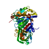
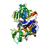
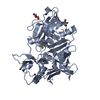
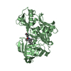
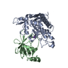
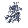
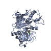
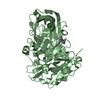
- PDB-2irz: Crystal structure of human Beta-secretase complexed with inhibitor -
+ Open data

- Basic information
| Entry | Database: PDB / ID: 2irz | ||||||
|---|---|---|---|---|---|---|---|
| Title | Crystal structure of human Beta-secretase complexed with inhibitor | ||||||
 Components Components | Beta-secretase 1 | ||||||
 Keywords Keywords | HYDROLASE / Aspartyl Protease | ||||||
| Function / homology |  Function and homology information Function and homology informationmemapsin 2 / Golgi-associated vesicle lumen / beta-aspartyl-peptidase activity / signaling receptor ligand precursor processing / amyloid-beta formation / amyloid precursor protein catabolic process / membrane protein ectodomain proteolysis / amyloid-beta metabolic process / detection of mechanical stimulus involved in sensory perception of pain / response to insulin-like growth factor stimulus ...memapsin 2 / Golgi-associated vesicle lumen / beta-aspartyl-peptidase activity / signaling receptor ligand precursor processing / amyloid-beta formation / amyloid precursor protein catabolic process / membrane protein ectodomain proteolysis / amyloid-beta metabolic process / detection of mechanical stimulus involved in sensory perception of pain / response to insulin-like growth factor stimulus / prepulse inhibition / cellular response to manganese ion / multivesicular body / swimming behavior / cellular response to copper ion / presynaptic modulation of chemical synaptic transmission / hippocampal mossy fiber to CA3 synapse / protein serine/threonine kinase binding / trans-Golgi network / protein processing / recycling endosome / response to lead ion / cellular response to amyloid-beta / synaptic vesicle / late endosome / peptidase activity / positive regulation of neuron apoptotic process / amyloid-beta binding / endopeptidase activity / amyloid fibril formation / aspartic-type endopeptidase activity / early endosome / lysosome / endosome / endosome membrane / membrane raft / endoplasmic reticulum lumen / Amyloid fiber formation / axon / neuronal cell body / dendrite / enzyme binding / cell surface / Golgi apparatus / proteolysis / membrane / plasma membrane Similarity search - Function | ||||||
| Biological species |  Homo sapiens (human) Homo sapiens (human) | ||||||
| Method |  X-RAY DIFFRACTION / MOLECULAR REPLACEMENT / Resolution: 1.8 Å X-RAY DIFFRACTION / MOLECULAR REPLACEMENT / Resolution: 1.8 Å | ||||||
 Authors Authors | Munshi, S. | ||||||
 Citation Citation | Journal: J.Med.Chem. / Year: 2006 Title: Discovery of oxadiazoyl tertiary carbinamine inhibitors of beta-secretase (BACE-1). Authors: Rajapakse, H.A. / Nantermet, P.G. / Selnick, H.G. / Munshi, S. / McGaughey, G.B. / Lindsley, S.R. / Young, M.B. / Lai, M.T. / Espeseth, A.S. / Shi, X.P. / Colussi, D. / Pietrak, B. / ...Authors: Rajapakse, H.A. / Nantermet, P.G. / Selnick, H.G. / Munshi, S. / McGaughey, G.B. / Lindsley, S.R. / Young, M.B. / Lai, M.T. / Espeseth, A.S. / Shi, X.P. / Colussi, D. / Pietrak, B. / Crouthamel, M.C. / Tugusheva, K. / Huang, Q. / Xu, M. / Simon, A.J. / Kuo, L. / Hazuda, D.J. / Graham, S. / Vacca, J.P. | ||||||
| History |
|
- Structure visualization
| Structure viewer | Molecule: MolmilJmol/JSmol |
|---|
- Downloads & links
-Download
| PDBx/mmCIF format | 2irz.cif.gz | 96.1 KB | Display | PDBx/mmCIF format |
|---|---|---|---|---|
| PDB format | pdb2irz.ent.gz | 71.6 KB | Display | PDB format |
| PDBx/mmJSON format | 2irz.json.gz | Tree view | PDBx/mmJSON format | |
| Others |  Other downloads Other downloads |
-Validation report
| Arichive directory | https://data.pdbj.org/pub/pdb/validation_reports/ir/2irzftp://data.pdbj.org/pub/pdb/validation_reports/ir/2irz | HTTPS FTP |
|---|
-Related structure data
| Related structure data | 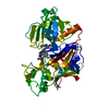 2is0C 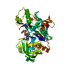 1tqfS S: Starting model for refinement C: citing same article ( |
|---|---|
| Similar structure data |


-Links
 PDBj
PDBj

- Assembly
| Deposited unit | 
| ||||||||
|---|---|---|---|---|---|---|---|---|---|
| 1 |
| ||||||||
| Unit cell |
|
-Components
| #1: Protein | Mass: 45122.750 Da / Num. of mol.: 1 / Fragment: protease domain / Mutation: K75A, E77A Source method: isolated from a genetically manipulated source Source: (gene. exp.) Homo sapiens (human) / Gene: BACE1, BACE / Plasmid: PET11A / Species (production host): Escherichia coli / Production host:  |
|---|---|
| #2: Chemical | ChemComp-I02 / 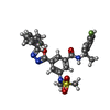  Mass: 551.632 Da / Num. of mol.: 1 / Source method: obtained synthetically / Formula: C28H30FN5O4S Mass: 551.632 Da / Num. of mol.: 1 / Source method: obtained synthetically / Formula: C28H30FN5O4S |
| #3: Water | ChemComp-HOH /  Mass: 18.015 Da / Num. of mol.: 294 / Source method: isolated from a natural source / Formula: H2O Mass: 18.015 Da / Num. of mol.: 294 / Source method: isolated from a natural source / Formula: H2O |
| Has protein modification | Y |
-Experimental details
-Experiment
| Experiment | Method: X-RAY DIFFRACTION / Number of used crystals: 1 |
|---|
- Sample preparation
| Crystal | Density Matthews: 2.83 Å3/Da / Density % sol: 56.49 % |
|---|---|
| Crystal grow | Temperature: 293 K / Method: vapor diffusion, sitting drop / pH: 7.5 Details: 1.5M Lithium sulfate, 0.1M HEPES buffer, pH 7.5, VAPOR DIFFUSION, SITTING DROP, temperature 293K |
-Data collection
| Diffraction | Mean temperature: 100 K |
|---|---|
| Diffraction source | Source: ROTATING ANODE / Type: RIGAKU RU300 / Wavelength: 1.5418 Å |
| Detector | Type: RIGAKU RAXIS IV / Detector: IMAGE PLATE / Date: Jan 24, 2004 |
| Radiation | Protocol: SINGLE WAVELENGTH / Monochromatic (M) / Laue (L): M / Scattering type: x-ray |
| Radiation wavelength | Wavelength: 1.5418 Å / Relative weight: 1 |
| Reflection | Resolution: 1.8→50 Å / Num. all: 45288 / Num. obs: 45288 / % possible obs: 94.8 % / Observed criterion σ(F): 0 / Observed criterion σ(I): 0 / Redundancy: 3 % / Rmerge(I) obs: 0.087 / Net I/σ(I): 23.5 |
| Reflection shell | Resolution: 1.8→1.86 Å / Redundancy: 2.9 % / Rmerge(I) obs: 0.585 / Mean I/σ(I) obs: 2 / Num. unique all: 4715 / % possible all: 96.8 |
- Processing
| Software |
| ||||||||||||||||||||
|---|---|---|---|---|---|---|---|---|---|---|---|---|---|---|---|---|---|---|---|---|---|
| Refinement | Method to determine structure: MOLECULAR REPLACEMENT Starting model: PDB entry 1TQF Resolution: 1.8→6 Å / σ(F): 0 / σ(I): 0
| ||||||||||||||||||||
| Refinement step | Cycle: LAST / Resolution: 1.8→6 Å
|
