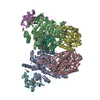
 Movie
Movie Controller


+ Open data

- Basic information
| Entry | Database: PDB / ID: 2inp | ||||||
|---|---|---|---|---|---|---|---|
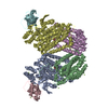
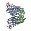
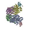
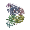
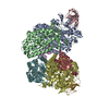
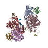
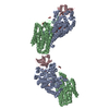
| Title | Structure of the Phenol Hydroxylase-Regulatory Protein Complex | ||||||
 Components Components | (Phenol hydroxylase component ...) x 4 | ||||||
 Keywords Keywords | OXIDOREDUCTASE / hydroxylase / diiron / four-helix bundle / regulatory protein | ||||||
| Function / homology |  Function and homology information Function and homology informationphenol 2-monooxygenase activity / oxidoreductase activity, acting on paired donors, with incorporation or reduction of molecular oxygen, NAD(P)H as one donor, and incorporation of one atom of oxygen / monooxygenase activity / metal ion binding Similarity search - Function | ||||||
| Biological species |  Pseudomonas stutzeri (bacteria) Pseudomonas stutzeri (bacteria) | ||||||
| Method |  X-RAY DIFFRACTION / SYNCHROTRON / MOLECULAR REPLACEMENT / Resolution: 2.3 Å X-RAY DIFFRACTION / SYNCHROTRON / MOLECULAR REPLACEMENT / Resolution: 2.3 Å | ||||||
 Authors Authors | Sazinsky, M.S. / Dunten, P.W. / McCormick, M.S. / Lippard, S.J. | ||||||
 Citation Citation | Journal: Biochemistry / Year: 2006 Title: X-ray Structure of a Hydroxylase-Regulatory Protein Complex from a Hydrocarbon-Oxidizing Multicomponent Monooxygenase, Pseudomonas sp. OX1 Phenol Hydroxylase. Authors: Sazinsky, M.H. / Dunten, P.W. / McCormick, M.S. / Didonato, A. / Lippard, S.J. | ||||||
| History |
|
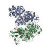
- Structure visualization
| Structure viewer | Molecule: MolmilJmol/JSmol |
|---|
- Downloads & links
-Download
| PDBx/mmCIF format | 2inp.cif.gz | 423.3 KB | Display | PDBx/mmCIF format |
|---|---|---|---|---|
| PDB format | pdb2inp.ent.gz | 342.4 KB | Display | PDB format |
| PDBx/mmJSON format | 2inp.json.gz | Tree view | PDBx/mmJSON format | |
| Others |  Other downloads Other downloads |
-Validation report
| Arichive directory | https://data.pdbj.org/pub/pdb/validation_reports/in/2inpftp://data.pdbj.org/pub/pdb/validation_reports/in/2inp | HTTPS FTP |
|---|
-Related structure data
| Related structure data |  2innSC S: Starting model for refinement C: citing same article ( |
|---|---|
| Similar structure data |
-Links
 PDBj
PDBj
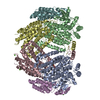
- Assembly
| Deposited unit | 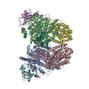
| ||||||||
|---|---|---|---|---|---|---|---|---|---|
| 1 |
| ||||||||
| Unit cell |
|
-Components
-Phenol hydroxylase component ... , 4 types, 7 molecules ABCDEFL
| #1: Protein | Mass: 58401.516 Da / Num. of mol.: 2 Source method: isolated from a genetically manipulated source Source: (gene. exp.) Pseudomonas stutzeri (bacteria) / Gene: phN / Production host: #2: Protein | Mass: 37968.840 Da / Num. of mol.: 2 Source method: isolated from a genetically manipulated source Source: (gene. exp.) Pseudomonas stutzeri (bacteria) / Gene: phL / Production host: #3: Protein | Mass: 13116.934 Da / Num. of mol.: 2 Source method: isolated from a genetically manipulated source Source: (gene. exp.) Pseudomonas stutzeri (bacteria) / Gene: phO / Production host: #4: Protein | | Mass: 10488.678 Da / Num. of mol.: 1 Source method: isolated from a genetically manipulated source Source: (gene. exp.) Pseudomonas stutzeri (bacteria) / Gene: phM / Production host: |
|---|
-Non-polymers , 3 types, 1028 molecules 


| #5: Chemical | ChemComp-FE /  Mass: 55.845 Da / Num. of mol.: 4 / Source method: obtained synthetically / Formula: Fe Mass: 55.845 Da / Num. of mol.: 4 / Source method: obtained synthetically / Formula: Fe#6: Chemical |  Mass: 65.409 Da / Num. of mol.: 2 / Source method: obtained synthetically / Formula: Zn Mass: 65.409 Da / Num. of mol.: 2 / Source method: obtained synthetically / Formula: Zn#7: Water | ChemComp-HOH / | Mass: 18.015 Da / Num. of mol.: 1022 / Source method: isolated from a natural source / Formula: H2O |
|---|
-Experimental details
-Experiment
| Experiment | Method: X-RAY DIFFRACTION / Number of used crystals: 1 |
|---|
- Sample preparation
| Crystal | Density Matthews: 2.66 Å3/Da / Density % sol: 53.71 % |
|---|---|
| Crystal grow | Temperature: 293 K / Method: vapor diffusion, hanging drop / pH: 7 Details: 100 mM Tris, pH 7.0, 150 mM Na2MoO4, 5% glycerol, and 17-20% PEG 8000 (w/w), VAPOR DIFFUSION, HANGING DROP, temperature 293K |
-Data collection
| Diffraction | Mean temperature: 100 K |
|---|---|
| Diffraction source | Source: SYNCHROTRON / Site: APS  / Beamline: 8-BM / Wavelength: 0.979 Å / Beamline: 8-BM / Wavelength: 0.979 Å |
| Detector | Type: ADSC QUANTUM 315 / Detector: CCD / Date: Jan 15, 2004 |
| Radiation | Monochromator: Si 111 CHANNEL / Protocol: SINGLE WAVELENGTH / Monochromatic (M) / Laue (L): M / Scattering type: x-ray |
| Radiation wavelength | Wavelength: 0.979 Å / Relative weight: 1 |
| Reflection | Resolution: 2.3→30 Å / Num. all: 108695 / Num. obs: 108695 / % possible obs: 100 % / Observed criterion σ(F): 2 / Observed criterion σ(I): 2 / Redundancy: 7.1 % / Rmerge(I) obs: 0.089 / Rsym value: 0.088 / Net I/σ(I): 16.5 |
| Reflection shell | Resolution: 2.3→2.36 Å / Redundancy: 6.9 % / Rmerge(I) obs: 0.448 / Mean I/σ(I) obs: 4.9 / Num. unique all: 7777 / Rsym value: 0.448 / % possible all: 100 |
- Processing
| Software |
| |||||||||||||||||||||||||||||||||||||||||||||||||||||||||||||||||||||||||||||||||||||||||||||||
|---|---|---|---|---|---|---|---|---|---|---|---|---|---|---|---|---|---|---|---|---|---|---|---|---|---|---|---|---|---|---|---|---|---|---|---|---|---|---|---|---|---|---|---|---|---|---|---|---|---|---|---|---|---|---|---|---|---|---|---|---|---|---|---|---|---|---|---|---|---|---|---|---|---|---|---|---|---|---|---|---|---|---|---|---|---|---|---|---|---|---|---|---|---|---|---|---|
| Refinement | Method to determine structure: MOLECULAR REPLACEMENT Starting model: PDB ENTRY 2INN Resolution: 2.3→30 Å / Cor.coef. Fo:Fc: 0.963 / Cor.coef. Fo:Fc free: 0.935 / SU B: 6.519 / SU ML: 0.157 / Cross valid method: THROUGHOUT / σ(F): 0 / ESU R: 0.281 / ESU R Free: 0.218 / Stereochemistry target values: MAXIMUM LIKELIHOOD
| |||||||||||||||||||||||||||||||||||||||||||||||||||||||||||||||||||||||||||||||||||||||||||||||
| Solvent computation | Ion probe radii: 0.8 Å / Shrinkage radii: 0.8 Å / VDW probe radii: 1.2 Å / Solvent model: MASK | |||||||||||||||||||||||||||||||||||||||||||||||||||||||||||||||||||||||||||||||||||||||||||||||
| Displacement parameters | Biso mean: 40.83 Å2
| |||||||||||||||||||||||||||||||||||||||||||||||||||||||||||||||||||||||||||||||||||||||||||||||
| Refinement step | Cycle: LAST / Resolution: 2.3→30 Å
| |||||||||||||||||||||||||||||||||||||||||||||||||||||||||||||||||||||||||||||||||||||||||||||||
| Refine LS restraints |
| |||||||||||||||||||||||||||||||||||||||||||||||||||||||||||||||||||||||||||||||||||||||||||||||
| LS refinement shell | Resolution: 2.302→2.362 Å / Total num. of bins used: 20
|
