 Movie
Movie Controller

[English] 日本語
 Yorodumi
- PDB-2ilu: Crystal structure of lactaldehyde dehydrogenase from E. coli: the... -
+ Open data

- Basic information
| Entry | Database: PDB / ID: 2ilu | ||||||
|---|---|---|---|---|---|---|---|
| Title | Crystal structure of lactaldehyde dehydrogenase from E. coli: the binary complex with NADPH | ||||||
 Components Components | Aldehyde dehydrogenase A | ||||||
 Keywords Keywords | OXIDOREDUCTASE / NADPH-Lactaldehyde dehydrogenase complex | ||||||
| Function / homology |  Function and homology information Function and homology informationglycolaldehyde dehydrogenase / glycolaldehyde dehydrogenase (NAD+) activity / lactaldehyde dehydrogenase / succinate-semialdehyde dehydrogenase (NAD+) activity / rhamnose catabolic process / lactaldehyde dehydrogenase (NAD+) activity / GABA catabolic process / L-fucose catabolic process / protein-containing complex / identical protein binding / cytosol Similarity search - Function | ||||||
| Biological species |  | ||||||
| Method |  X-RAY DIFFRACTION / SYNCHROTRON / FOURIER SYNTHESIS / Resolution: 2.7 Å X-RAY DIFFRACTION / SYNCHROTRON / FOURIER SYNTHESIS / Resolution: 2.7 Å | ||||||
 Authors Authors | Di Costanzo, L. / Gomez, G. / Christianson, D.W. | ||||||
 Citation Citation | Journal: J.Mol.Biol. / Year: 2007 Title: Crystal structure of lactaldehyde dehydrogenase from Escherichia coli and inferences regarding substrate and cofactor specificity. Authors: Di Costanzo, L. / Gomez, G.A. / Christianson, D.W. | ||||||
| History |
|
- Structure visualization
| Structure viewer | Molecule: MolmilJmol/JSmol |
|---|
- Downloads & links
-Download
| PDBx/mmCIF format | 2ilu.cif.gz | 105.2 KB | Display | PDBx/mmCIF format |
|---|---|---|---|---|
| PDB format | pdb2ilu.ent.gz | 80.5 KB | Display | PDB format |
| PDBx/mmJSON format | 2ilu.json.gz | Tree view | PDBx/mmJSON format | |
| Others |  Other downloads Other downloads |
-Validation report
| Arichive directory | https://data.pdbj.org/pub/pdb/validation_reports/il/2iluftp://data.pdbj.org/pub/pdb/validation_reports/il/2ilu | HTTPS FTP |
|---|
-Related structure data
| Related structure data |  2hg2SC 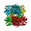 2impC S: Starting model for refinement C: citing same article ( |
|---|---|
| Similar structure data |


-Links
 PDBj
PDBj


- Assembly
| Deposited unit | 
| ||||||||
|---|---|---|---|---|---|---|---|---|---|
| 1 | 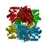
| ||||||||
| Unit cell |
| ||||||||
| Details | The biological assembly is a tetramer generated from the monomer in the asymmetric unit by the operations: -x, -y, z (4); y, x, -z+1/3 (7); -y, -x, -z+1/3 (10) |
-Components
| #1: Protein | Mass: 52372.527 Da / Num. of mol.: 1 Source method: isolated from a genetically manipulated source Source: (gene. exp.) References: UniProt: P25553, lactaldehyde dehydrogenase, glycolaldehyde dehydrogenase | ||||
|---|---|---|---|---|---|
| #2: Chemical |   Mass: 96.063 Da / Num. of mol.: 2 / Source method: obtained synthetically / Formula: SO4 Mass: 96.063 Da / Num. of mol.: 2 / Source method: obtained synthetically / Formula: SO4#3: Chemical | ChemComp-NDP / |   Mass: 745.421 Da / Num. of mol.: 1 / Source method: obtained synthetically / Formula: C21H30N7O17P3 Mass: 745.421 Da / Num. of mol.: 1 / Source method: obtained synthetically / Formula: C21H30N7O17P3#4: Water | ChemComp-HOH / |  Mass: 18.015 Da / Num. of mol.: 32 / Source method: isolated from a natural source / Formula: H2O Mass: 18.015 Da / Num. of mol.: 32 / Source method: isolated from a natural source / Formula: H2O |
-Experimental details
-Experiment
| Experiment | Method: X-RAY DIFFRACTION / Number of used crystals: 1 |
|---|
- Sample preparation
| Crystal | Density Matthews: 3.09 Å3/Da / Density % sol: 60.17 % |
|---|---|
| Crystal grow | Temperature: 298 K / Method: vapor diffusion, hanging drop Details: 0.1 M Bis-Tris or Tris, 2.0 M ammonium sulfate, pH pH 6.8 - 7.2, VAPOR DIFFUSION, HANGING DROP, temperature 298K |
-Data collection
| Diffraction | Mean temperature: 100 K |
|---|---|
| Diffraction source | Source: SYNCHROTRON / Site: CHESS  / Beamline: F1 / Wavelength: 0.9798 Å / Beamline: F1 / Wavelength: 0.9798 Å |
| Detector | Type: ADSC QUANTUM 4 / Detector: CCD / Date: Feb 5, 2006 |
| Radiation | Protocol: SINGLE WAVELENGTH / Monochromatic (M) / Laue (L): M / Scattering type: x-ray |
| Radiation wavelength | Wavelength: 0.9798 Å / Relative weight: 1 |
| Reflection | Resolution: 2.7→50 Å / Num. obs: 16757 / Redundancy: 14.4 % / Rmerge(I) obs: 0.139 / Net I/σ(I): 22.6 |
- Processing
| Software |
| ||||||||||||||||
|---|---|---|---|---|---|---|---|---|---|---|---|---|---|---|---|---|---|
| Refinement | Method to determine structure: FOURIER SYNTHESIS Starting model: 2HG2 Resolution: 2.7→50 Å
| ||||||||||||||||
| Refinement step | Cycle: LAST / Resolution: 2.7→50 Å
| ||||||||||||||||
| Refine LS restraints |
| ||||||||||||||||
| LS refinement shell | Resolution: 2.7→2.8 Å / Rfactor Rfree: 0.2945 / Rfactor Rwork: 0.2506 |
