 Movie
Movie Controller

[English] 日本語
 Yorodumi
- PDB-2ikq: Crystal structure of mouse Sts-1 PGM domain in complex with phosphate -
+ Open data

- Basic information
| Entry | Database: PDB / ID: 2ikq | ||||||
|---|---|---|---|---|---|---|---|
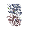
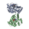
| Title | Crystal structure of mouse Sts-1 PGM domain in complex with phosphate | ||||||
 Components Components | Suppressor of T-cell receptor signaling 1 | ||||||
 Keywords Keywords | SIGNALING PROTEIN / IMMUNE SYSTEM / PGM / acid phosphatase / phospho-histidine enzyme | ||||||
| Function / homology |  Function and homology information Function and homology informationcollagen-activated tyrosine kinase receptor signaling pathway / collagen-activated signaling pathway / regulation of osteoclast differentiation / negative regulation of platelet aggregation / regulation of release of sequestered calcium ion into cytosol / negative regulation of bone resorption / negative regulation of osteoclast differentiation / negative regulation of signal transduction / protein-tyrosine-phosphatase / protein tyrosine phosphatase activity ...collagen-activated tyrosine kinase receptor signaling pathway / collagen-activated signaling pathway / regulation of osteoclast differentiation / negative regulation of platelet aggregation / regulation of release of sequestered calcium ion into cytosol / negative regulation of bone resorption / negative regulation of osteoclast differentiation / negative regulation of signal transduction / protein-tyrosine-phosphatase / protein tyrosine phosphatase activity / phosphoprotein binding / platelet activation / platelet aggregation / ubiquitin protein ligase binding / identical protein binding / nucleus / cytoplasm Similarity search - Function | ||||||
| Biological species |  | ||||||
| Method |  X-RAY DIFFRACTION / SYNCHROTRON / FOURIER SYNTHESIS / Resolution: 2.609 Å X-RAY DIFFRACTION / SYNCHROTRON / FOURIER SYNTHESIS / Resolution: 2.609 Å | ||||||
 Authors Authors | Chen, Y. / Nassar, N. | ||||||
 Citation Citation | Journal: Mol.Cell / Year: 2007 Title: A Phosphatase Activity of Sts-1 Contributes to the Suppression of TCR Signaling Authors: Mikhailik, A. / Ford, B. / Keller, J. / Chen, Y. / Nassar, N. / Carpino, N. | ||||||
| History |
|
- Structure visualization
| Structure viewer | Molecule: MolmilJmol/JSmol |
|---|
- Downloads & links
-Download
| PDBx/mmCIF format | 2ikq.cif.gz | 159.4 KB | Display | PDBx/mmCIF format |
|---|---|---|---|---|
| PDB format | pdb2ikq.ent.gz | 126.5 KB | Display | PDB format |
| PDBx/mmJSON format | 2ikq.json.gz | Tree view | PDBx/mmJSON format | |
| Others |  Other downloads Other downloads |
-Validation report
| Arichive directory | https://data.pdbj.org/pub/pdb/validation_reports/ik/2ikqftp://data.pdbj.org/pub/pdb/validation_reports/ik/2ikq | HTTPS FTP |
|---|
-Related structure data
| Related structure data |  2h0qC  1h0qS C: citing same article ( S: Starting model for refinement |
|---|---|
| Similar structure data |


-Links
 PDBj
PDBj


- Assembly
| Deposited unit | 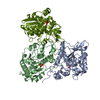
| ||||||||||||||||||||||||||||||||||||||||||||||||||||||||||||||||||||||||||||||||||||||||||||||||||||||||||||||||||||||||||||||||||||||||||||||||||||||||||||||||||||||||||||||||||||||||||||||||||||||||||||||||
|---|---|---|---|---|---|---|---|---|---|---|---|---|---|---|---|---|---|---|---|---|---|---|---|---|---|---|---|---|---|---|---|---|---|---|---|---|---|---|---|---|---|---|---|---|---|---|---|---|---|---|---|---|---|---|---|---|---|---|---|---|---|---|---|---|---|---|---|---|---|---|---|---|---|---|---|---|---|---|---|---|---|---|---|---|---|---|---|---|---|---|---|---|---|---|---|---|---|---|---|---|---|---|---|---|---|---|---|---|---|---|---|---|---|---|---|---|---|---|---|---|---|---|---|---|---|---|---|---|---|---|---|---|---|---|---|---|---|---|---|---|---|---|---|---|---|---|---|---|---|---|---|---|---|---|---|---|---|---|---|---|---|---|---|---|---|---|---|---|---|---|---|---|---|---|---|---|---|---|---|---|---|---|---|---|---|---|---|---|---|---|---|---|---|---|---|---|---|---|---|---|---|---|---|---|---|---|---|---|---|
| 1 | 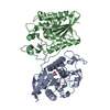
| ||||||||||||||||||||||||||||||||||||||||||||||||||||||||||||||||||||||||||||||||||||||||||||||||||||||||||||||||||||||||||||||||||||||||||||||||||||||||||||||||||||||||||||||||||||||||||||||||||||||||||||||||
| 2 | 
| ||||||||||||||||||||||||||||||||||||||||||||||||||||||||||||||||||||||||||||||||||||||||||||||||||||||||||||||||||||||||||||||||||||||||||||||||||||||||||||||||||||||||||||||||||||||||||||||||||||||||||||||||
| Unit cell |
| ||||||||||||||||||||||||||||||||||||||||||||||||||||||||||||||||||||||||||||||||||||||||||||||||||||||||||||||||||||||||||||||||||||||||||||||||||||||||||||||||||||||||||||||||||||||||||||||||||||||||||||||||
| Noncrystallographic symmetry (NCS) | NCS domain:
NCS domain segments:
|