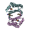
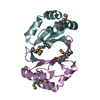
- PDB-2ifx: Crystal structure of a putative 4-methylmuconolactone methylisome... -
+
Open data
ID or keywords:
Loading...
-
Basic information
Entry
Database: PDB / ID: 2ifx
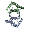
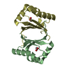
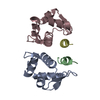
Title
Crystal structure of a putative 4-methylmuconolactone methylisomerase (YP_295714.1) from Ralstonia eutropha JMP134 at 2.00 A resolution
Components
Hypothetical protein
Keywords
Structural Genomics/Unknown function / YP_295714.1 / hypothetical protein / Structural Genomics / PSI-2 / Protein Structure Initiative / Joint Center for Structural Genomics / JCSG / Structural Genomics-Unknown function COMPLEX
Function / homology
Function and homology information
4-carboxymethyl-4-methylbutenolide mutase / 4-carboxymethyl-4-methylbutenolide mutase activity Similarity search - Function
Resolution: 2→25.743 Å / Num. obs: 17384 / % possible obs: 94.7 % / Biso Wilson estimate: 44.084 Å2 / Rmerge(I) obs: 0.045 / Net I/σ(I): 10.34
Reflection shell
Diffraction-ID: 1
Resolution (Å)
Highest resolution (Å)
Rmerge(I) obs
Mean I/σ(I) obs
Num. measured obs
Num. unique all
% possible all
2-2.07
0.328
2.5
5095
2702
80
2.07-2.15
0.225
3.6
5934
3089
91.5
2.15-2.25
0.161
4.6
6610
3423
94.8
2.25-2.37
0.132
5.6
6466
3354
95.9
2.37-2.52
0.094
7.4
6764
3457
97.1
2.52-2.71
0.076
9.7
6446
3355
98
2.71-2.99
0.056
12.7
6739
3475
98.4
2.99-3.42
0.042
16.2
6603
3415
98.9
3.42
0.043
18.9
6560
3425
98.4
-
Phasing
Phasing
Method: MAD
-
Processing
Software
Name
Version
Classification
NB
MolProbity
3beta29
modelbuilding
REFMAC
5.2.0019
refinement
XSCALE
datascaling
PDB_EXTRACT
2
dataextraction
XDS
datareduction
SOLVE
phasing
RESOLVE
phasing
Refinement
Method to determine structure: MAD / Resolution: 2→25.743 Å / Cor.coef. Fo:Fc: 0.967 / Cor.coef. Fo:Fc free: 0.955 / SU B: 7.935 / SU ML: 0.108 / TLS residual ADP flag: LIKELY RESIDUAL / Cross valid method: THROUGHOUT / σ(F): 0 / ESU R: 0.155 / ESU R Free: 0.137 Stereochemistry target values: MAXIMUM LIKELIHOOD WITH PHASES Details: 1. HYDROGENS HAVE BEEN ADDED IN THE RIDING POSITIONS. 2. A MET-INHIBITION PROTOCOL WAS USED FOR SELENOMETHIONINE INCORPORATION DURING PROTEIN EXPRESSION. THE OCCUPANCY OF THE SE ATOMS IN THE ...Details: 1. HYDROGENS HAVE BEEN ADDED IN THE RIDING POSITIONS. 2. A MET-INHIBITION PROTOCOL WAS USED FOR SELENOMETHIONINE INCORPORATION DURING PROTEIN EXPRESSION. THE OCCUPANCY OF THE SE ATOMS IN THE MSE RESIDUES WAS REDUCED TO 0.75 FOR THE REDUCED SCATTERING POWER DUE TO PARTIAL S-MET INCORPORATION. 3. RESIDUES 49-51, 109-113 IN CHAIN A, AND 49-59, 108-113 IN CHAIN B ARE DISORDERED AND NOT INCLUDED IN THE MODEL. 4. GLYCEROL MOLECULES FROM THE CRYO SOLUTION ARE MODELED. 5. ATOM RECORDS CONTAIN RESIDUAL B FACTORS ONLY. 6. THERE ARE SOME UNEXPLAINED DENSITY NEAR RESIDUES B15(SER) AND B39(TYR). 7. SIGNIFICANT NCS DEVIATION OCCURS IN THE RESIDUE RANGE 83-100.
Rfactor
Num. reflection
% reflection
Selection details
Rfree
0.202
882
5.1 %
RANDOM
Rwork
0.171
-
-
-
obs
0.173
17337
99.29 %
-
Solvent computation
Ion probe radii: 0.8 Å / Shrinkage radii: 0.8 Å / VDW probe radii: 1.2 Å / Solvent model: BABINET MODEL WITH MASK
In the structure databanks used in Yorodumi, some data are registered as the other names, "COVID-19 virus" and "2019-nCoV". Here are the details of the virus and the list of structure data.
Jan 31, 2019. EMDB accession codes are about to change! (news from PDBe EMDB page)
EMDB accession codes are about to change! (news from PDBe EMDB page)
The allocation of 4 digits for EMDB accession codes will soon come to an end. Whilst these codes will remain in use, new EMDB accession codes will include an additional digit and will expand incrementally as the available range of codes is exhausted. The current 4-digit format prefixed with “EMD-” (i.e. EMD-XXXX) will advance to a 5-digit format (i.e. EMD-XXXXX), and so on. It is currently estimated that the 4-digit codes will be depleted around Spring 2019, at which point the 5-digit format will come into force.
The EM Navigator/Yorodumi systems omit the EMD- prefix.
Related info.:Q: What is EMD? / ID/Accession-code notation in Yorodumi/EM Navigator
Yorodumi is a browser for structure data from EMDB, PDB, SASBDB, etc.
This page is also the successor to EM Navigator detail page, and also detail information page/front-end page for Omokage search.
The word "yorodu" (or yorozu) is an old Japanese word meaning "ten thousand". "mi" (miru) is to see.
Related info.:EMDB / PDB / SASBDB / Comparison of 3 databanks / Yorodumi Search / Aug 31, 2016. New EM Navigator & Yorodumi / Yorodumi Papers / Jmol/JSmol / Function and homology information / Changes in new EM Navigator and Yorodumi
 Movie
Movie Controller

 Yorodumi
 Open data
 Basic information
 Components
 Keywords
 Function and homology information
 Cupriavidus necator (bacteria)
 X-RAY DIFFRACTION /
  Authors
 Citation
 Structure visualization
 Downloads & links
 Other downloads
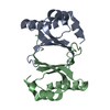


 PDBj
PDBj Assembly
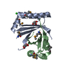

 Mass: 35.453 Da / Num. of mol.: 1 / Source method: obtained synthetically / Formula: Cl
 Mass: 92.094 Da / Num. of mol.: 2 / Source method: obtained synthetically / Formula: C3H8O3
 Mass: 18.015 Da / Num. of mol.: 125 / Source method: isolated from a natural source / Formula: H2O
 Sample preparation
 / Beamline: 23-ID-D / Wavelength: 0.94926, 0.97925, 0.97939
 Processing