 Movie
Movie Controller

[English] 日本語
 Yorodumi
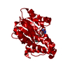
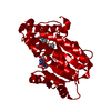
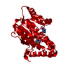
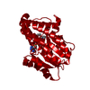
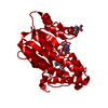
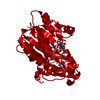
- PDB-2idz: Crystal structure of wild type Enoyl-ACP(CoA) reductase from Myco... -
+ Open data

- Basic information
| Entry | Database: PDB / ID: 2idz | ||||||
|---|---|---|---|---|---|---|---|
| Title | Crystal structure of wild type Enoyl-ACP(CoA) reductase from Mycobacterium tuberculosis in complex with NADH-INH | ||||||
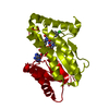
 Components Components | Enoyl-[acyl-carrier-protein] reductase [NADH] | ||||||
 Keywords Keywords | OXIDOREDUCTASE / ENOYL-ACYL CARRIER PROTEIN / InhA / INH / isoniazid | ||||||
| Function / homology |  Function and homology information Function and homology informationenoyl-[acyl-carrier-protein] reductase [NAD(P)H] activity / trans-2-enoyl-CoA reductase (NADH) activity / mycolic acid biosynthetic process / fatty acid elongation / enoyl-[acyl-carrier-protein] reductase (NADH) / enoyl-[acyl-carrier-protein] reductase (NADH) activity / NAD+ binding / peptidoglycan-based cell wall / fatty acid binding / fatty acid biosynthetic process ...enoyl-[acyl-carrier-protein] reductase [NAD(P)H] activity / trans-2-enoyl-CoA reductase (NADH) activity / mycolic acid biosynthetic process / fatty acid elongation / enoyl-[acyl-carrier-protein] reductase (NADH) / enoyl-[acyl-carrier-protein] reductase (NADH) activity / NAD+ binding / peptidoglycan-based cell wall / fatty acid binding / fatty acid biosynthetic process / response to antibiotic / plasma membrane Similarity search - Function | ||||||
| Biological species |   Mycobacterium tuberculosis (bacteria) Mycobacterium tuberculosis (bacteria) | ||||||
| Method |  X-RAY DIFFRACTION / SYNCHROTRON / MOLECULAR REPLACEMENT / Resolution: 2 Å X-RAY DIFFRACTION / SYNCHROTRON / MOLECULAR REPLACEMENT / Resolution: 2 Å | ||||||
 Authors Authors | Dias, M.V.B. / Prado, M.P.X. / Vasconcelos, I.B. / Valmir, F. / Basso, L.A. / Santos, D.S. / Azevedo Jr., W.F. | ||||||
 Citation Citation | Journal: J.Struct.Biol. / Year: 2007 Title: Crystallographic studies on the binding of isonicotinyl-NAD adduct to wild-type and isoniazid resistant 2-trans-enoyl-ACP (CoA) reductase from Mycobacterium tuberculosis. Authors: Dias, M.V. / Vasconcelos, I.B. / Prado, A.M. / Fadel, V. / Basso, L.A. / de Azevedo, W.F. / Santos, D.S. | ||||||
| History |
|
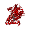
- Structure visualization
| Structure viewer | Molecule: MolmilJmol/JSmol |
|---|
- Downloads & links
-Download
| PDBx/mmCIF format | 2idz.cif.gz | 71.9 KB | Display | PDBx/mmCIF format |
|---|---|---|---|---|
| PDB format | pdb2idz.ent.gz | 52.6 KB | Display | PDB format |
| PDBx/mmJSON format | 2idz.json.gz | Tree view | PDBx/mmJSON format | |
| Others |  Other downloads Other downloads |
-Validation report
| Arichive directory | https://data.pdbj.org/pub/pdb/validation_reports/id/2idzftp://data.pdbj.org/pub/pdb/validation_reports/id/2idz | HTTPS FTP |
|---|
-Related structure data
| Related structure data |  2ie0C  2iebC 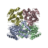 2iedC  1zidS C: citing same article ( S: Starting model for refinement |
|---|---|
| Similar structure data |


-Links
 PDBj
PDBj

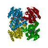
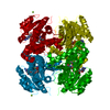
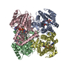
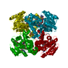
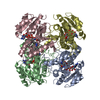
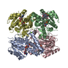
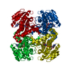
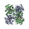
- Assembly
| Deposited unit | 
| ||||||||
|---|---|---|---|---|---|---|---|---|---|
| 1 | 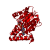
| ||||||||
| Unit cell |
| ||||||||
| Components on special symmetry positions |
|
-Components
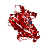
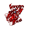
| #1: Protein | Mass: 28423.586 Da / Num. of mol.: 1 Source method: isolated from a genetically manipulated source Source: (gene. exp.) Mycobacterium tuberculosis (bacteria) / Gene: inhA / Plasmid: PET-23D(+) / Species (production host): Escherichia coli / Production host: References: UniProt: P0A5Y6, UniProt: P9WGR1*PLUS, enoyl-[acyl-carrier-protein] reductase (NADH) |
|---|---|
| #2: Chemical | ChemComp-ZID /   Mass: 768.519 Da / Num. of mol.: 1 / Source method: obtained synthetically / Formula: C27H30N8O15P2 Mass: 768.519 Da / Num. of mol.: 1 / Source method: obtained synthetically / Formula: C27H30N8O15P2 |
| #3: Water | ChemComp-HOH /  Mass: 18.015 Da / Num. of mol.: 237 / Source method: isolated from a natural source / Formula: H2O Mass: 18.015 Da / Num. of mol.: 237 / Source method: isolated from a natural source / Formula: H2O |
-Experimental details
-Experiment
| Experiment | Method: X-RAY DIFFRACTION / Number of used crystals: 1 |
|---|
- Sample preparation
| Crystal | Density Matthews: 3.28 Å3/Da / Density % sol: 62.5 % |
|---|---|
| Crystal grow | Temperature: 293 K / Method: vapor diffusion, hanging drop / pH: 7.2 Details: 0.05M SODIUM CITRATE, 0.05M HEPES, 8-15% 2-METHYL-2-4-PENTANEDIOL (MPD), PH 7.2, TEMPERATURE 293K , VAPOR DIFFUSION, HANGING DROP |
-Data collection
| Diffraction | Mean temperature: 100 K |
|---|---|
| Diffraction source | Source: SYNCHROTRON / Site: LNLS  / Beamline: D03B-MX1 / Wavelength: 1.427 Å / Beamline: D03B-MX1 / Wavelength: 1.427 Å |
| Detector | Type: MAR CCD 130 mm / Detector: CCD / Date: Mar 22, 2006 |
| Radiation | Monochromator: GRAPHITE / Protocol: SINGLE WAVELENGTH / Monochromatic (M) / Laue (L): M / Scattering type: x-ray |
| Radiation wavelength | Wavelength: 1.427 Å / Relative weight: 1 |
| Reflection | Resolution: 2→40.193 Å / Num. obs: 26409 / % possible obs: 96.11 % / Observed criterion σ(F): 2 / Observed criterion σ(I): 2 / Rmerge(I) obs: 0.098 / Rsym value: 0.094 / Net I/σ(I): 5.2 |
| Reflection shell | Resolution: 2→2.11 Å / Rmerge(I) obs: 0.83 / Mean I/σ(I) obs: 2.3 / Num. unique all: 3795 / % possible all: 100 |
- Processing
| Software |
| ||||||||||||||||||||||||||||||||||||||||||||||||||||||||||||||||||||||||||||||||||||||||||
|---|---|---|---|---|---|---|---|---|---|---|---|---|---|---|---|---|---|---|---|---|---|---|---|---|---|---|---|---|---|---|---|---|---|---|---|---|---|---|---|---|---|---|---|---|---|---|---|---|---|---|---|---|---|---|---|---|---|---|---|---|---|---|---|---|---|---|---|---|---|---|---|---|---|---|---|---|---|---|---|---|---|---|---|---|---|---|---|---|---|---|---|
| Refinement | Method to determine structure: MOLECULAR REPLACEMENT Starting model: 1ZID Resolution: 2→40.19 Å / Cor.coef. Fo:Fc: 0.96 / Cor.coef. Fo:Fc free: 0.945 / SU B: 3.5 / SU ML: 0.098 / Cross valid method: THROUGHOUT / σ(F): 2 / ESU R: 0.145 / ESU R Free: 0.139 / Stereochemistry target values: MAXIMUM LIKELIHOOD / Details: HYDROGENS HAVE BEEN ADDED IN THE RIDING POSITIONS
| ||||||||||||||||||||||||||||||||||||||||||||||||||||||||||||||||||||||||||||||||||||||||||
| Solvent computation | Ion probe radii: 0.8 Å / Shrinkage radii: 0.8 Å / VDW probe radii: 1.2 Å / Solvent model: MASK | ||||||||||||||||||||||||||||||||||||||||||||||||||||||||||||||||||||||||||||||||||||||||||
| Displacement parameters | Biso mean: 32.205 Å2
| ||||||||||||||||||||||||||||||||||||||||||||||||||||||||||||||||||||||||||||||||||||||||||
| Refinement step | Cycle: LAST / Resolution: 2→40.19 Å
| ||||||||||||||||||||||||||||||||||||||||||||||||||||||||||||||||||||||||||||||||||||||||||
| Refine LS restraints |
| ||||||||||||||||||||||||||||||||||||||||||||||||||||||||||||||||||||||||||||||||||||||||||
| LS refinement shell | Resolution: 2→2.052 Å
|
