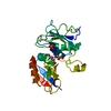
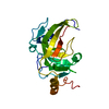
BIOMOLECULE: 1 THIS ENTRY CONTAINS THE CRYSTALLOGRAPHIC ASYMMETRIC UNIT WHICH CONSISTS OF 1 ... BIOMOLECULE: 1 THIS ENTRY CONTAINS THE CRYSTALLOGRAPHIC ASYMMETRIC UNIT WHICH CONSISTS OF 1 CHAIN(S). SEE REMARK 350 FOR INFORMATION ON GENERATING THE BIOLOGICAL MOLECULE(S). SIZE EXCLUSION CHROMATOGRAPHY SUPPORTS THE ASSIGNMENT OF A MONOMER AS A BIOLOGICALLY SIGNIFICANT OLIGOMERIZATION STATE.
Resolution: 1.75→29.501 Å / Num. obs: 19754 / % possible obs: 93.9 % / Biso Wilson estimate: 27.791 Å2 / Rmerge(I) obs: 0.056 / Net I/σ(I): 9.19
Reflection shell
Diffraction-ID: 1
Resolution (Å)
Highest resolution (Å)
Rmerge(I) obs
Mean I/σ(I) obs
Num. measured obs
Num. unique all
% possible all
1.75-1.81
0.361
2.2
5713
3015
83.8
1.81-1.89
0.303
2.7
6838
3605
88
1.89-1.97
0.229
3.5
5933
3130
90.2
1.97-2.07
0.172
4.4
6339
3340
93.6
2.07-2.2
0.121
6.2
6776
3590
95.5
2.2-2.37
0.097
7.6
6831
3618
96.8
2.37-2.61
0.07
9.8
6949
3664
97.1
2.61-2.99
0.059
13.1
6896
3662
97.6
2.99
0.043
17.6
6813
3650
98.3
-
Phasing
Phasing
Method: MAD
-
Processing
Software
Name
Version
Classification
NB
MolProbity
3beta29
modelbuilding
SHELX
phasing
REFMAC
5.2.0019
refinement
XSCALE
datascaling
PDB_EXTRACT
2
dataextraction
XDS
datareduction
SHELXD
phasing
SHARP
phasing
Refinement
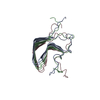
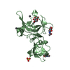
Method to determine structure: MAD / Resolution: 1.75→29.501 Å / Cor.coef. Fo:Fc: 0.952 / Cor.coef. Fo:Fc free: 0.931 / WRfactor Rwork: 0.194 / SU B: 4.648 / SU ML: 0.083 / TLS residual ADP flag: LIKELY RESIDUAL / Cross valid method: THROUGHOUT / σ(F): 0 / ESU R: 0.119 / ESU R Free: 0.121 Stereochemistry target values: MAXIMUM LIKELIHOOD WITH PHASES Details: (1) HYDROGENS HAVE BEEN ADDED IN THE RIDING POSITIONS. (2) ZN200 IS COORDINATED TO THE SIDE CHAINS OF CYS 8, 10, 28 AND 29. ZN201 IS COORDINATED TO THE SIDE CHAINS OF CYS 167, 169, 178 AND ...Details: (1) HYDROGENS HAVE BEEN ADDED IN THE RIDING POSITIONS. (2) ZN200 IS COORDINATED TO THE SIDE CHAINS OF CYS 8, 10, 28 AND 29. ZN201 IS COORDINATED TO THE SIDE CHAINS OF CYS 167, 169, 178 AND 179. ZN IS MODELED BASED ON ELECTRON DENSITY AND GEOMETRY, AND IS SUPPORTED BY X-RAY FLOURESCENCE EXPERIMENTS. (3) A MET-INHIBITION PROTOCOL WAS USED FOR SELENOMETHIONINE INCORPORATION DURING PROTEIN EXPRESSION. THE OCCUPANCY OF THE SE ATOMS IN THE MSE RESIDUES WAS REDUCED TO 0.75 TO ACCOUNT FOR THE REDUCED SCATTERING POWER DUE TO PARTIAL S-MET INCORPORATION. (4) ATOM RECORD CONTAINS RESIDUAL B FACTORS ONLY. (5) THE RESIDUES IN THE DISORDERED REGIONS OF A40-45 WERE NOT MODELLED. (6) THERE ARE UNMODELED DENSITIES NEAR A132 AND A56.
Rfactor
Num. reflection
% reflection
Selection details
Rfree
0.238
1007
5.1 %
RANDOM
Rwork
0.192
-
-
-
obs
0.19392
19707
99.23 %
-
Solvent computation
Ion probe radii: 0.8 Å / Shrinkage radii: 0.8 Å / VDW probe radii: 1.2 Å / Solvent model: MASK
Displacement parameters
Biso mean: 20.135 Å2
Baniso -1
Baniso -2
Baniso -3
1-
1.6 Å2
0 Å2
0 Å2
2-
-
-0.26 Å2
0 Å2
3-
-
-
-1.34 Å2
Refinement step
Cycle: LAST / Resolution: 1.75→29.501 Å
Protein
Nucleic acid
Ligand
Solvent
Total
Num. atoms
1370
0
3
127
1500
Refine LS restraints
Refine-ID
Type
Dev ideal
Dev ideal target
Number
X-RAY DIFFRACTION
r_bond_refined_d
0.015
0.022
1429
X-RAY DIFFRACTION
r_bond_other_d
0.001
0.02
940
X-RAY DIFFRACTION
r_angle_refined_deg
1.522
1.943
1950
X-RAY DIFFRACTION
r_angle_other_deg
0.903
3
2307
X-RAY DIFFRACTION
r_dihedral_angle_1_deg
5.811
5
180
X-RAY DIFFRACTION
r_dihedral_angle_2_deg
37.61
24.068
59
X-RAY DIFFRACTION
r_dihedral_angle_3_deg
13.377
15
230
X-RAY DIFFRACTION
r_dihedral_angle_4_deg
7.59
15
5
X-RAY DIFFRACTION
r_chiral_restr
0.087
0.2
220
X-RAY DIFFRACTION
r_gen_planes_refined
0.006
0.02
1586
X-RAY DIFFRACTION
r_gen_planes_other
0.001
0.02
284
X-RAY DIFFRACTION
r_nbd_refined
0.235
0.2
249
X-RAY DIFFRACTION
r_nbd_other
0.189
0.2
905
X-RAY DIFFRACTION
r_nbtor_refined
0.181
0.2
680
X-RAY DIFFRACTION
r_nbtor_other
0.087
0.2
699
X-RAY DIFFRACTION
r_xyhbond_nbd_refined
0.131
0.2
79
X-RAY DIFFRACTION
r_symmetry_vdw_refined
0.289
0.2
18
X-RAY DIFFRACTION
r_symmetry_vdw_other
0.269
0.2
34
X-RAY DIFFRACTION
r_symmetry_hbond_refined
0.125
0.2
7
X-RAY DIFFRACTION
r_mcbond_it
2.096
3
967
X-RAY DIFFRACTION
r_mcbond_other
0.505
3
352
X-RAY DIFFRACTION
r_mcangle_it
2.978
5
1454
X-RAY DIFFRACTION
r_scbond_it
4.796
8
593
X-RAY DIFFRACTION
r_scangle_it
6.423
11
493
LS refinement shell
Resolution: 1.751→1.797 Å / Total num. of bins used: 20
Rfactor
Num. reflection
% reflection
Rfree
0.287
68
-
Rwork
0.236
1350
-
obs
-
1418
98.88 %
Refinement TLS params.
Method: refined / Refine-ID: X-RAY DIFFRACTION
ID
L11 (°2)
L12 (°2)
L13 (°2)
L22 (°2)
L23 (°2)
L33 (°2)
S11 (Å °)
S12 (Å °)
S13 (Å °)
S21 (Å °)
S22 (Å °)
S23 (Å °)
S31 (Å °)
S32 (Å °)
S33 (Å °)
T11 (Å2)
T12 (Å2)
T13 (Å2)
T22 (Å2)
T23 (Å2)
T33 (Å2)
Origin x (Å)
Origin y (Å)
Origin z (Å)
1
0.7903
-0.332
-0.2126
3.24
0.3876
3.0368
0.1472
0.0039
-0.1117
0.1111
-0.0837
-0.2089
0.246
-0.1805
-0.0635
-0.0468
-0.0608
-0.0285
-0.0294
0.0255
-0.0195
11.8959
11.0248
22.1427
2
1.1208
-0.291
0.5634
2.2314
-0.3807
3.1129
0.0897
0.0164
0.0223
-0.0065
-0.0886
-0.2232
-0.0951
0.0708
-0.0012
-0.1175
-0.0064
-0.0072
-0.0676
0.0207
-0.0286
14.6595
23.6128
16.2532
3
2.3253
0.6989
4.4883
3.3019
-0.2022
13.6996
0.0529
0.16
-0.0098
0.0529
0.0097
-0.1044
-0.3371
0.3062
-0.0626
-0.1549
0.0155
0.0079
-0.0413
0.0135
-0.0551
11.5322
28.4246
17.252
4
1.5283
-0.366
-0.0553
1.6295
-1.6871
4.8341
-0.0293
0.0605
0.0431
-0.0723
-0.0305
-0.0006
-0.6592
0.0035
0.0598
0.0722
0.0175
-0.0015
-0.0821
0.0152
-0.0789
10.5745
35.1568
3.586
Refinement TLS group
Refine-ID: X-RAY DIFFRACTION / Selection: ALL / Auth asym-ID: A / Label asym-ID: A
ID
Refine TLS-ID
Auth seq-ID
Label seq-ID
1
1
1 - 39
2 - 40
2
2
46 - 123
47 - 124
3
3
127 - 143
128 - 144
4
4
145 - 183
146 - 184
+
About Yorodumi
-
News
-
Feb 9, 2022. New format data for meta-information of EMDB entries
New format data for meta-information of EMDB entries
Version 3 of the EMDB header file is now the official format.
The previous official version 1.9 will be removed from the archive.
In the structure databanks used in Yorodumi, some data are registered as the other names, "COVID-19 virus" and "2019-nCoV". Here are the details of the virus and the list of structure data.
Jan 31, 2019. EMDB accession codes are about to change! (news from PDBe EMDB page)
EMDB accession codes are about to change! (news from PDBe EMDB page)
The allocation of 4 digits for EMDB accession codes will soon come to an end. Whilst these codes will remain in use, new EMDB accession codes will include an additional digit and will expand incrementally as the available range of codes is exhausted. The current 4-digit format prefixed with “EMD-” (i.e. EMD-XXXX) will advance to a 5-digit format (i.e. EMD-XXXXX), and so on. It is currently estimated that the 4-digit codes will be depleted around Spring 2019, at which point the 5-digit format will come into force.
The EM Navigator/Yorodumi systems omit the EMD- prefix.
Related info.:Q: What is EMD? / ID/Accession-code notation in Yorodumi/EM Navigator
Yorodumi is a browser for structure data from EMDB, PDB, SASBDB, etc.
This page is also the successor to EM Navigator detail page, and also detail information page/front-end page for Omokage search.
The word "yorodu" (or yorozu) is an old Japanese word meaning "ten thousand". "mi" (miru) is to see.
Related info.:EMDB / PDB / SASBDB / Comparison of 3 databanks / Yorodumi Search / Aug 31, 2016. New EM Navigator & Yorodumi / Yorodumi Papers / Jmol/JSmol / Function and homology information / Changes in new EM Navigator and Yorodumi
 Movie
Movie Controller

 Yorodumi
 Open data
 Basic information
 Components
 Keywords
 Function and homology information
 Psychrobacter arcticus (bacteria)
 X-RAY DIFFRACTION /
  Authors
 Citation
 Structure visualization
 Downloads & links
 Other downloads
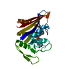


 PDBj
PDBj
 Assembly

 Mass: 65.409 Da / Num. of mol.: 2 / Source method: obtained synthetically / Formula: Zn
 Mass: 35.453 Da / Num. of mol.: 1 / Source method: obtained synthetically / Formula: Cl
 Mass: 18.015 Da / Num. of mol.: 127 / Source method: isolated from a natural source / Formula: H2O
 Sample preparation
 / Beamline: 23-ID-D / Wavelength: 0.94926,0.97939,0.97925
 Processing