 Movie
Movie Controller

[English] 日本語
 Yorodumi
- PDB-2i9l: Structure of Fab 7D11 from a neutralizing antibody against the po... -
+ Open data

- Basic information
| Entry | Database: PDB / ID: 2i9l | ||||||
|---|---|---|---|---|---|---|---|
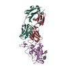
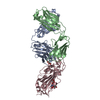
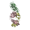
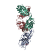
| Title | Structure of Fab 7D11 from a neutralizing antibody against the poxvirus L1 protein | ||||||
 Components Components |
| ||||||
 Keywords Keywords | IMMUNE SYSTEM/VIRAL PROTEIN / Neutralizing antibody / Poxvirus / antibody complex / IMMUNE SYSTEM-VIRAL PROTEIN COMPLEX | ||||||
| Function / homology |  Function and homology information Function and homology informationviral envelope / symbiont entry into host cell / virion attachment to host cell / virion membrane / membrane Similarity search - Function | ||||||
| Biological species |  Vaccinia virus Vaccinia virus | ||||||
| Method |  X-RAY DIFFRACTION / SYNCHROTRON / MOLECULAR REPLACEMENT / Resolution: 3.1 Å X-RAY DIFFRACTION / SYNCHROTRON / MOLECULAR REPLACEMENT / Resolution: 3.1 Å | ||||||
 Authors Authors | Su, H.P. / Golden, J.W. / Gittis, A.G. / Moss, B. / Hooper, J.W. / Garboczi, D.N. | ||||||
 Citation Citation | Journal: Virology / Year: 2007 Title: Structural basis for the binding of the neutralizing antibody, 7D11, to the poxvirus L1 protein Authors: Su, H.-P. / Golden, J.W. / Gittis, A.G. / Hooper, J.W. / Garboczi, D.N. | ||||||
| History |
|
- Structure visualization
| Structure viewer | Molecule: MolmilJmol/JSmol |
|---|
- Downloads & links
-Download
| PDBx/mmCIF format | 2i9l.cif.gz | 459.5 KB | Display | PDBx/mmCIF format |
|---|---|---|---|---|
| PDB format | pdb2i9l.ent.gz | 377.6 KB | Display | PDB format |
| PDBx/mmJSON format | 2i9l.json.gz | Tree view | PDBx/mmJSON format | |
| Others |  Other downloads Other downloads |
-Validation report
| Summary document | 2i9l_validation.pdf.gz | 530.7 KB | Display | wwPDB validaton report |
|---|---|---|---|---|
| Full document | 2i9l_full_validation.pdf.gz | 610.6 KB | Display | |
| Data in XML | 2i9l_validation.xml.gz | 90.7 KB | Display | |
| Data in CIF | 2i9l_validation.cif.gz | 122.1 KB | Display | |
| Arichive directory | https://data.pdbj.org/pub/pdb/validation_reports/i9/2i9lftp://data.pdbj.org/pub/pdb/validation_reports/i9/2i9l | HTTPS FTP |
-Related structure data


-Links
 PDBj
PDBj


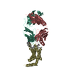
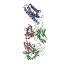
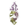
- Assembly
| Deposited unit | 
| ||||||||
|---|---|---|---|---|---|---|---|---|---|
| 1 | 
| ||||||||
| 2 | 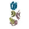
| ||||||||
| 3 | 
| ||||||||
| 4 | 
| ||||||||
| Unit cell |
|
-Components
| #1: Antibody | Mass: 24175.852 Da / Num. of mol.: 4 / Source method: isolated from a natural source / Details: Antibody light chain isolated from a hybridoma / Source: (natural) #2: Antibody | Mass: 23668.525 Da / Num. of mol.: 4 / Source method: isolated from a natural source / Details: Antibody heavy chain isolated from a hybridoma / Source: (natural) #3: Protein | Mass: 19532.877 Da / Num. of mol.: 4 Source method: isolated from a genetically manipulated source Source: (gene. exp.) Vaccinia virus / Genus: Orthopoxvirus / Strain: Western Reserve / Gene: l1r / Plasmid: pNAN / Production host:  #4: Chemical | ChemComp-GOL /   Mass: 92.094 Da / Num. of mol.: 8 / Source method: obtained synthetically / Formula: C3H8O3 Mass: 92.094 Da / Num. of mol.: 8 / Source method: obtained synthetically / Formula: C3H8O3#5: Water | ChemComp-HOH / |  Mass: 18.015 Da / Num. of mol.: 14 / Source method: isolated from a natural source / Formula: H2O Mass: 18.015 Da / Num. of mol.: 14 / Source method: isolated from a natural source / Formula: H2OHas protein modification | Y | |
|---|
-Experimental details
-Experiment
| Experiment | Method: X-RAY DIFFRACTION / Number of used crystals: 1 |
|---|
- Sample preparation
| Crystal | Density Matthews: 3.2 Å3/Da / Density % sol: 61.53 % |
|---|---|
| Crystal grow | Temperature: 298 K / pH: 8 Details: 10% PEG 10000, 0.1M Tris, VAPOR DIFFUSION, HANGING DROP, temperature 298K, pH 8.00 |
-Data collection
| Diffraction | Mean temperature: 100 K |
|---|---|
| Diffraction source | Source: SYNCHROTRON / Site: APS  / Beamline: 19-ID / Wavelength: 0.97915 / Beamline: 19-ID / Wavelength: 0.97915 |
| Detector | Type: ADSC QUANTUM 315 / Detector: CCD / Date: Nov 14, 2005 |
| Radiation | Monochromator: SI / Protocol: SINGLE WAVELENGTH / Monochromatic (M) / Laue (L): M / Scattering type: x-ray |
| Radiation wavelength | Wavelength: 0.97915 Å / Relative weight: 1 |
| Reflection | Resolution: 3.1→50 Å / Num. obs: 61289 / % possible obs: 98.6 % / Redundancy: 3.3 % / Rmerge(I) obs: 0.124 / Net I/σ(I): 7 |
| Reflection shell | Resolution: 3.1→3.21 Å / Redundancy: 3.2 % / Rmerge(I) obs: 0.464 / % possible all: 99.6 |
-Phasing
| Phasing MR |
|
|---|
- Processing
| Software |
| ||||||||||||||||||||||||||||||||||||||||||||||||||||||||||||||||||||||||||||||||
|---|---|---|---|---|---|---|---|---|---|---|---|---|---|---|---|---|---|---|---|---|---|---|---|---|---|---|---|---|---|---|---|---|---|---|---|---|---|---|---|---|---|---|---|---|---|---|---|---|---|---|---|---|---|---|---|---|---|---|---|---|---|---|---|---|---|---|---|---|---|---|---|---|---|---|---|---|---|---|---|---|---|
| Refinement | Method to determine structure: MOLECULAR REPLACEMENT Starting model: 1YPY, 1YMH Resolution: 3.1→50 Å / Isotropic thermal model: ISOTROPIC / Cross valid method: THROUGHOUT / σ(F): 0 / Stereochemistry target values: ENGH & HUBER
| ||||||||||||||||||||||||||||||||||||||||||||||||||||||||||||||||||||||||||||||||
| Solvent computation | Bsol: 10 Å2 | ||||||||||||||||||||||||||||||||||||||||||||||||||||||||||||||||||||||||||||||||
| Displacement parameters | Biso mean: 39.19 Å2 | ||||||||||||||||||||||||||||||||||||||||||||||||||||||||||||||||||||||||||||||||
| Refinement step | Cycle: LAST / Resolution: 3.1→50 Å
| ||||||||||||||||||||||||||||||||||||||||||||||||||||||||||||||||||||||||||||||||
| Refine LS restraints |
| ||||||||||||||||||||||||||||||||||||||||||||||||||||||||||||||||||||||||||||||||
| LS refinement shell | Resolution: 3.1→3.12 Å / Total num. of bins used: 50 /
| ||||||||||||||||||||||||||||||||||||||||||||||||||||||||||||||||||||||||||||||||
| Xplor file |
|
