 Movie
Movie Controller

[English] 日本語
 Yorodumi
- PDB-2i0z: Crystal structure of a FAD binding protein from Bacillus cereus, ... -
+ Open data

- Basic information
| Entry | Database: PDB / ID: 2i0z | ||||||
|---|---|---|---|---|---|---|---|
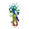
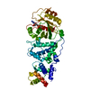
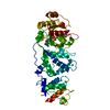
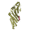
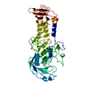
| Title | Crystal structure of a FAD binding protein from Bacillus cereus, a putative NAD(FAD)-utilizing dehydrogenases | ||||||
 Components Components | NAD(FAD)-utilizing dehydrogenases | ||||||
 Keywords Keywords | OXIDOREDUCTASE / Structural Genomics / NAD(FAD)-utilizing dehydrogenases / PSI-2 / Protein Structure Initiative / Midwest Center for Structural Genomics / MCSG | ||||||
| Function / homology |  Function and homology information Function and homology information | ||||||
| Biological species |  | ||||||
| Method |  X-RAY DIFFRACTION / SYNCHROTRON / MOLECULAR REPLACEMENT / Resolution: 1.84 Å X-RAY DIFFRACTION / SYNCHROTRON / MOLECULAR REPLACEMENT / Resolution: 1.84 Å | ||||||
 Authors Authors | Minasov, G. / Shuvalova, L. / Vorontsov, I.I. / Kiryukhina, O. / Abdullah, J. / Collart, F.R. / Joachimiak, A. / Anderson, W.F. / Midwest Center for Structural Genomics (MCSG) | ||||||
 Citation Citation | Journal: To be Published Title: Crystal structure of a FAD binding protein from Bacillus cereus, a putative NAD(FAD)-utilizing dehydrogenases Authors: Minasov, G. / Shuvalova, L. / Vorontsov, I.I. / Kiryukhina, O. / Abdullah, J. / Collart, F.R. / Joachimiak, A. / Anderson, W.F. | ||||||
| History |
|
- Structure visualization
| Structure viewer | Molecule: MolmilJmol/JSmol |
|---|
- Downloads & links
-Download
| PDBx/mmCIF format | 2i0z.cif.gz | 108.3 KB | Display | PDBx/mmCIF format |
|---|---|---|---|---|
| PDB format | pdb2i0z.ent.gz | 79.7 KB | Display | PDB format |
| PDBx/mmJSON format | 2i0z.json.gz | Tree view | PDBx/mmJSON format | |
| Others |  Other downloads Other downloads |
-Validation report
| Arichive directory | https://data.pdbj.org/pub/pdb/validation_reports/i0/2i0zftp://data.pdbj.org/pub/pdb/validation_reports/i0/2i0z | HTTPS FTP |
|---|
-Related structure data
| Related structure data |  2gqfS S: Starting model for refinement |
|---|---|
| Similar structure data | |
| Other databases |
-Links
 PDBj
PDBj- Assembly
| Deposited unit | 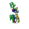
| ||||||||
|---|---|---|---|---|---|---|---|---|---|
| 1 |
| ||||||||
| Unit cell |
| ||||||||
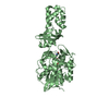
| Details | Biological unit is a monomer |
-Components
| #1: Protein | Mass: 49012.848 Da / Num. of mol.: 1 / Mutation: P99H Source method: isolated from a genetically manipulated source Source: (gene. exp.) |
|---|---|
| #2: Chemical | ChemComp-CL /   Mass: 35.453 Da / Num. of mol.: 1 / Source method: obtained synthetically / Formula: Cl Mass: 35.453 Da / Num. of mol.: 1 / Source method: obtained synthetically / Formula: Cl |
| #3: Chemical | ChemComp-FAD /   Mass: 785.550 Da / Num. of mol.: 1 / Source method: obtained synthetically / Formula: C27H33N9O15P2 / Comment: FAD*YM Mass: 785.550 Da / Num. of mol.: 1 / Source method: obtained synthetically / Formula: C27H33N9O15P2 / Comment: FAD*YM |
| #4: Water | ChemComp-HOH /  Mass: 18.015 Da / Num. of mol.: 373 / Source method: isolated from a natural source / Formula: H2O Mass: 18.015 Da / Num. of mol.: 373 / Source method: isolated from a natural source / Formula: H2O |
-Experimental details
-Experiment
| Experiment | Method: X-RAY DIFFRACTION / Number of used crystals: 1 |
|---|
- Sample preparation
| Crystal | Density Matthews: 2.15 Å3/Da / Density % sol: 42.67 % |
|---|---|
| Crystal grow | Temperature: 295 K / Method: vapor diffusion, sitting drop / pH: 8.3 Details: 0.01M TRIS-HCL, 0.2M Ammonium chloride, 0.25M Sodium chloride, 20% PEG 3350, pH 8.3, VAPOR DIFFUSION, SITTING DROP, temperature 295K |
-Data collection
| Diffraction | Mean temperature: 100 K |
|---|---|
| Diffraction source | Source: SYNCHROTRON / Site: APS  / Beamline: 23-ID-D / Wavelength: 0.984 Å / Beamline: 23-ID-D / Wavelength: 0.984 Å |
| Detector | Type: MARRESEARCH / Detector: CCD / Date: Mar 6, 2006 / Details: mirrors |
| Radiation | Monochromator: Crystal / Protocol: SINGLE WAVELENGTH / Monochromatic (M) / Laue (L): M / Scattering type: x-ray |
| Radiation wavelength | Wavelength: 0.984 Å / Relative weight: 1 |
| Reflection | Resolution: 1.84→25 Å / Num. all: 36967 / Num. obs: 36967 / % possible obs: 98.9 % / Observed criterion σ(I): -3 / Redundancy: 3.9 % / Rmerge(I) obs: 0.075 / Net I/σ(I): 13.9 |
| Reflection shell | Resolution: 1.84→1.92 Å / Redundancy: 3.8 % / Rmerge(I) obs: 0.394 / Mean I/σ(I) obs: 3.9 / Num. unique all: 3617 / % possible all: 99.7 |
- Processing
| Software |
| ||||||||||||||||||||||||||||||||||||||||||||||||||||||||||||||||||||||||||||||||||||||||||
|---|---|---|---|---|---|---|---|---|---|---|---|---|---|---|---|---|---|---|---|---|---|---|---|---|---|---|---|---|---|---|---|---|---|---|---|---|---|---|---|---|---|---|---|---|---|---|---|---|---|---|---|---|---|---|---|---|---|---|---|---|---|---|---|---|---|---|---|---|---|---|---|---|---|---|---|---|---|---|---|---|---|---|---|---|---|---|---|---|---|---|---|
| Refinement | Method to determine structure: MOLECULAR REPLACEMENT Starting model: PDB ENTRY 2GQF Resolution: 1.84→24.86 Å / Cor.coef. Fo:Fc: 0.957 / Cor.coef. Fo:Fc free: 0.926 / SU B: 3.237 / SU ML: 0.102 Isotropic thermal model: Isotropic individual atomic B-factors refined Cross valid method: THROUGHOUT / σ(I): 0 / ESU R: 0.157 / ESU R Free: 0.152 / Stereochemistry target values: MAXIMUM LIKELIHOOD / Details: HYDROGENS HAVE BEEN ADDED IN THE RIDING POSITIONS
| ||||||||||||||||||||||||||||||||||||||||||||||||||||||||||||||||||||||||||||||||||||||||||
| Solvent computation | Ion probe radii: 0.8 Å / Shrinkage radii: 0.8 Å / VDW probe radii: 1.4 Å / Solvent model: BABINET MODEL WITH MASK | ||||||||||||||||||||||||||||||||||||||||||||||||||||||||||||||||||||||||||||||||||||||||||
| Displacement parameters | Biso mean: 34.978 Å2
| ||||||||||||||||||||||||||||||||||||||||||||||||||||||||||||||||||||||||||||||||||||||||||
| Refinement step | Cycle: LAST / Resolution: 1.84→24.86 Å
| ||||||||||||||||||||||||||||||||||||||||||||||||||||||||||||||||||||||||||||||||||||||||||
| Refine LS restraints |
| ||||||||||||||||||||||||||||||||||||||||||||||||||||||||||||||||||||||||||||||||||||||||||
| LS refinement shell | Resolution: 1.84→1.89 Å / Total num. of bins used: 20
|
