SEQUENCE THE CONSTRUCT WAS EXPRESSED WITH A PURIFICATION TAG MGSDKIHHHHHHENLYFQG. THE TAG WAS ...SEQUENCE THE CONSTRUCT WAS EXPRESSED WITH A PURIFICATION TAG MGSDKIHHHHHHENLYFQG. THE TAG WAS REMOVED WITH TEV PROTEASE LEAVING ONLY A GLYCINE (0) FOLLOWED BY THE TARGET SEQUENCE.
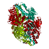
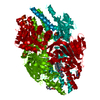
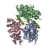
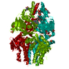
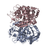
A: Predicted sugar phosphatases of the HAD superfamily B: Predicted sugar phosphatases of the HAD superfamily C: Predicted sugar phosphatases of the HAD superfamily D: Predicted sugar phosphatases of the HAD superfamily hetero molecules
Resolution: 2.1→29.323 Å / Num. obs: 70192 / % possible obs: 100 % / Redundancy: 3.7 % / Biso Wilson estimate: 20.6 Å2 / Rmerge(I) obs: 0.152 / Rsym value: 0.152 / Net I/σ(I): 4.4
Reflection shell
Resolution (Å)
Redundancy (%)
Rmerge(I) obs
Mean I/σ(I) obs
Num. measured all
Num. unique all
Rsym value
% possible all
2.1-2.15
3.7
0.635
1.2
18914
5087
0.635
100
2.15-2.21
3.7
0.546
1.4
18642
5016
0.546
100
2.21-2.28
3.7
0.5
1.2
18069
4877
0.5
100
2.28-2.35
3.7
0.445
1.7
17544
4714
0.445
100
2.35-2.42
3.7
0.394
1.9
17148
4593
0.394
100
2.42-2.51
3.7
0.34
2.1
16514
4437
0.34
100
2.51-2.6
3.7
0.308
2.5
16004
4296
0.308
100
2.6-2.71
3.7
0.264
2.8
15361
4126
0.264
100
2.71-2.83
3.7
0.228
3.3
14764
3970
0.228
100
2.83-2.97
3.7
0.172
4.4
14176
3814
0.172
100
2.97-3.13
3.7
0.13
5.6
13489
3629
0.13
100
3.13-3.32
3.7
0.11
6.5
12709
3429
0.11
100
3.32-3.55
3.7
0.093
6.9
11920
3218
0.093
100
3.55-3.83
3.7
0.082
8.1
11306
3054
0.082
100
3.83-4.2
3.7
0.07
8.1
10212
2789
0.07
100
4.2-4.7
3.7
0.063
9.4
9385
2556
0.063
100
4.7-5.42
3.6
0.065
9.5
8186
2258
0.065
100
5.42-6.64
3.6
0.077
8.4
6930
1937
0.077
100
6.64-9.39
3.5
0.062
9
5323
1525
0.062
99.9
9.39-29.32
3.2
0.045
11.1
2772
865
0.045
96.6
-
Phasing
Phasing
Method: MAD
-
Processing
Software
Name
Version
Classification
NB
MolProbity
3beta29
modelbuilding
SHELX
phasing
REFMAC
5.2.0019
refinement
SCALA
datascaling
PDB_EXTRACT
2
dataextraction
MOSFLM
datareduction
CCP4
(SCALA)
datascaling
SHELXD
phasing
autoSHARP
phasing
Refinement
Method to determine structure: MAD / Resolution: 2.1→29.323 Å / Cor.coef. Fo:Fc: 0.962 / Cor.coef. Fo:Fc free: 0.932 / SU B: 5.401 / SU ML: 0.142 / Cross valid method: THROUGHOUT / σ(F): 0 / ESU R: 0.236 / ESU R Free: 0.194 Stereochemistry target values: MAXIMUM LIKELIHOOD WITH PHASES Details: 1. HYDROGENS HAVE BEEN ADDED IN THE RIDING POSITIONS 2. NCS RESTRAINTS ARE APPLIED SUCH CHAIN A IS VERY SIMILAR TO CHAIN B, CHAIN C SIMILAR TO CHAIN D. THE NCS BREAKS DOWN FOR C108-129/D108- ...Details: 1. HYDROGENS HAVE BEEN ADDED IN THE RIDING POSITIONS 2. NCS RESTRAINTS ARE APPLIED SUCH CHAIN A IS VERY SIMILAR TO CHAIN B, CHAIN C SIMILAR TO CHAIN D. THE NCS BREAKS DOWN FOR C108-129/D108-129. 3. EACH ACTIVE SITE HAS A MAGNESIUM BOUND. MAGNESIUM IS PRESENT IN THE CYSTALLIZATION SOLUTION. BASED ON DIFFERENCE MAP, THERE APPEARS ANOTHER NON-WATER ENTITY (NEAR THR 52 OG1) FOR EACH MONOMER. THEY ARE TENATIVELY ASSIGNED AS CHLORIDES (21,22,23,24). A HEPES MOLECULE (PRESENT IN CRYSTALLIZATION SOLUTION) IS MODELLED INTO THE ACTIVE SITE. SINCE HEPES IS SIMILAR IN STRUCTURE TO SUBSTRATES OF THIS ENZYME CONTAINING RIBOSE-PHOSPHATE, IT COULD BE AN INHIBITOR. HOWEVER, DUE TO THE POOR DENSITY, THE ASSIGNMENT IS ONLY TENATIVE. 4. A MET-INHIBITION PROTOCOL WAS USED FOR SELENOMETHIONINE INCORPORATION DURING PROTEIN EXPRESSION. THE OCCUPANCY OF THE SE ATOMS IN THE MSE RESIDUES WAS REDUCED TO 0.7 TO ACCOUNT FOR THE REDUCED SCATTERING POWER DUE TO PARTIAL S-MET INCORPORATION.
Rfactor
Num. reflection
% reflection
Selection details
Rfree
0.234
3531
5 %
RANDOM
Rwork
0.179
-
-
-
all
0.181
-
-
-
obs
0.18146
70121
99.92 %
-
Solvent computation
Ion probe radii: 0.8 Å / Shrinkage radii: 0.8 Å / VDW probe radii: 1.2 Å / Solvent model: BABINET MODEL WITH MASK
Displacement parameters
Biso mean: 23.291 Å2
Baniso -1
Baniso -2
Baniso -3
1-
-0.59 Å2
0 Å2
0 Å2
2-
-
0.53 Å2
0 Å2
3-
-
-
0.06 Å2
Refinement step
Cycle: LAST / Resolution: 2.1→29.323 Å
Protein
Nucleic acid
Ligand
Solvent
Total
Num. atoms
8884
0
138
1035
10057
Refine LS restraints
Refine-ID
Type
Dev ideal
Dev ideal target
Number
X-RAY DIFFRACTION
r_bond_refined_d
0.016
0.022
9166
X-RAY DIFFRACTION
r_bond_other_d
0.002
0.02
6147
X-RAY DIFFRACTION
r_angle_refined_deg
1.433
1.981
12415
X-RAY DIFFRACTION
r_angle_other_deg
0.972
3
15150
X-RAY DIFFRACTION
r_dihedral_angle_1_deg
6.433
5
1186
X-RAY DIFFRACTION
r_dihedral_angle_2_deg
38.842
25.094
371
X-RAY DIFFRACTION
r_dihedral_angle_3_deg
13.952
15
1629
X-RAY DIFFRACTION
r_dihedral_angle_4_deg
15.681
15
35
X-RAY DIFFRACTION
r_chiral_restr
0.082
0.2
1451
X-RAY DIFFRACTION
r_gen_planes_refined
0.005
0.02
10069
X-RAY DIFFRACTION
r_gen_planes_other
0.002
0.02
1750
X-RAY DIFFRACTION
r_nbd_refined
0.209
0.2
1797
X-RAY DIFFRACTION
r_nbd_other
0.187
0.2
6453
X-RAY DIFFRACTION
r_nbtor_refined
0.179
0.2
4529
X-RAY DIFFRACTION
r_nbtor_other
0.086
0.2
4679
X-RAY DIFFRACTION
r_xyhbond_nbd_refined
0.181
0.2
791
X-RAY DIFFRACTION
r_metal_ion_refined
0.029
0.2
5
X-RAY DIFFRACTION
r_symmetry_vdw_refined
0.162
0.2
24
X-RAY DIFFRACTION
r_symmetry_vdw_other
0.246
0.2
46
X-RAY DIFFRACTION
r_symmetry_hbond_refined
0.161
0.2
26
X-RAY DIFFRACTION
r_mcbond_it
1.837
3
5878
X-RAY DIFFRACTION
r_mcbond_other
0.383
3
2356
X-RAY DIFFRACTION
r_mcangle_it
2.842
5
9257
X-RAY DIFFRACTION
r_scbond_it
5.194
8
3721
X-RAY DIFFRACTION
r_scangle_it
7.049
11
3129
Refine LS restraints NCS
Dom-ID: 1 / Refine-ID: X-RAY DIFFRACTION
Ens-ID
Auth asym-ID
Number
Type
Rms dev position (Å)
Weight position
1
A
1640
TIGHTPOSITIONAL
0.05
0.05
1
A
1905
MEDIUMPOSITIONAL
0.28
0.5
1
A
1640
TIGHTTHERMAL
0.17
0.5
1
A
1905
MEDIUMTHERMAL
1.01
2
2
C
1512
TIGHTPOSITIONAL
0.07
0.05
2
C
1898
MEDIUMPOSITIONAL
0.29
0.5
2
C
125
LOOSEPOSITIONAL
0.98
5
2
C
1512
TIGHTTHERMAL
0.16
0.5
2
C
1898
MEDIUMTHERMAL
0.97
2
2
C
125
LOOSETHERMAL
3.98
10
LS refinement shell
Resolution: 2.1→2.155 Å / Total num. of bins used: 20
Rfactor
Num. reflection
% reflection
Rfree
0.296
245
-
Rwork
0.233
4828
-
obs
-
5073
99.74 %
+
About Yorodumi
-
News
-
Feb 9, 2022. New format data for meta-information of EMDB entries
New format data for meta-information of EMDB entries
Version 3 of the EMDB header file is now the official format.
The previous official version 1.9 will be removed from the archive.
In the structure databanks used in Yorodumi, some data are registered as the other names, "COVID-19 virus" and "2019-nCoV". Here are the details of the virus and the list of structure data.
Jan 31, 2019. EMDB accession codes are about to change! (news from PDBe EMDB page)
EMDB accession codes are about to change! (news from PDBe EMDB page)
The allocation of 4 digits for EMDB accession codes will soon come to an end. Whilst these codes will remain in use, new EMDB accession codes will include an additional digit and will expand incrementally as the available range of codes is exhausted. The current 4-digit format prefixed with “EMD-” (i.e. EMD-XXXX) will advance to a 5-digit format (i.e. EMD-XXXXX), and so on. It is currently estimated that the 4-digit codes will be depleted around Spring 2019, at which point the 5-digit format will come into force.
The EM Navigator/Yorodumi systems omit the EMD- prefix.
Related info.:Q: What is EMD? / ID/Accession-code notation in Yorodumi/EM Navigator
Yorodumi is a browser for structure data from EMDB, PDB, SASBDB, etc.
This page is also the successor to EM Navigator detail page, and also detail information page/front-end page for Omokage search.
The word "yorodu" (or yorozu) is an old Japanese word meaning "ten thousand". "mi" (miru) is to see.
Related info.:EMDB / PDB / SASBDB / Comparison of 3 databanks / Yorodumi Search / Aug 31, 2016. New EM Navigator & Yorodumi / Yorodumi Papers / Jmol/JSmol / Function and homology information / Changes in new EM Navigator and Yorodumi
 Movie
Movie Controller

 Yorodumi
 Open data
 Basic information
 Components
 Keywords
 Function and homology information
 Cytophaga hutchinsonii (bacteria)
 X-RAY DIFFRACTION /
  Authors
 Citation
 Structure visualization
 Downloads & links
 Other downloads
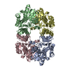


 PDBj
PDBj Assembly
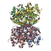


 Mass: 24.305 Da / Num. of mol.: 4 / Source method: obtained synthetically / Formula: Mg
 Mass: 35.453 Da / Num. of mol.: 15 / Source method: obtained synthetically / Formula: Cl
 Mass: 238.305 Da / Num. of mol.: 4 / Source method: obtained synthetically / Formula: C8H18N2O4S / Comment: pH buffer*YM
 Mass: 62.068 Da / Num. of mol.: 15 / Source method: obtained synthetically / Formula: C2H6O2
 Sample preparation
 / Beamline: BL9-2 / Wavelength: 0.97932, 0.91837, 0.97915
 Processing