 Movie
Movie Controller

[English] 日本語
 Yorodumi
- PDB-3poc: The crystal structure of the D307A mutant of alpha-Glucosidase (F... -
+ Open data

- Basic information
| Entry | Database: PDB / ID: 3poc | ||||||||||||
|---|---|---|---|---|---|---|---|---|---|---|---|---|---|
| Title | The crystal structure of the D307A mutant of alpha-Glucosidase (FAMILY 31) from Ruminococcus obeum ATCC 29174 in complex with acarbose | ||||||||||||
 Components Components | alpha-Glucosidase | ||||||||||||
 Keywords Keywords | hydrolase/hydrolase inhibitor / structural genomics / PSI-2 / protein structure initiative / midwest center for structural genomics / MCSG / multi-domain / alpha-Glucosidase / cytoplasmic / hydrolase-hydrolase inhibitor complex | ||||||||||||
| Function / homology |  Function and homology information Function and homology informationHydrolases; Glycosylases; Glycosidases, i.e. enzymes that hydrolyse O- and S-glycosyl compounds / hydrolase activity, hydrolyzing O-glycosyl compounds / carbohydrate binding / carbohydrate metabolic process Similarity search - Function | ||||||||||||
| Biological species |  Ruminococcus obeum (bacteria) Ruminococcus obeum (bacteria) | ||||||||||||
| Method |  X-RAY DIFFRACTION / SYNCHROTRON / MOLECULAR REPLACEMENT / Resolution: 1.99 Å X-RAY DIFFRACTION / SYNCHROTRON / MOLECULAR REPLACEMENT / Resolution: 1.99 Å | ||||||||||||
 Authors Authors | Tan, K. / Tesar, C. / Wilton, R. / Keigher, L. / Babnigg, G. / Joachimiak, A. / Midwest Center for Structural Genomics (MCSG) | ||||||||||||
 Citation Citation | Journal: To be Published Title: The crystal structure of the D307A mutant of alpha-Glucosidase (FAMILY 31) from Ruminococcus obeum ATCC 29174 in complex with acarbose Authors: Tan, K. / Tesar, C. / Wilton, R. / Keigher, L. / Babnigg, G. / Joachimiak, A. | ||||||||||||
| History |
|
- Structure visualization
| Structure viewer | Molecule: MolmilJmol/JSmol |
|---|
- Downloads & links
-Download
| PDBx/mmCIF format | 3poc.cif.gz | 555.7 KB | Display | PDBx/mmCIF format |
|---|---|---|---|---|
| PDB format | pdb3poc.ent.gz | 456 KB | Display | PDB format |
| PDBx/mmJSON format | 3poc.json.gz | Tree view | PDBx/mmJSON format | |
| Others |  Other downloads Other downloads |
-Validation report
| Arichive directory | https://data.pdbj.org/pub/pdb/validation_reports/po/3pocftp://data.pdbj.org/pub/pdb/validation_reports/po/3poc | HTTPS FTP |
|---|
-Related structure data
| Related structure data |  3n04S S: Starting model for refinement |
|---|---|
| Similar structure data | |
| Other databases |


-Links
 PDBj
PDBj


- Assembly
| Deposited unit | 
| ||||||||
|---|---|---|---|---|---|---|---|---|---|
| 1 |
| ||||||||
| Unit cell |
| ||||||||
| Details | The chains A and B form a dimer. |
-Components
-Protein , 1 types, 2 molecules AB
| #1: Protein | Mass: 77405.453 Da / Num. of mol.: 2 / Mutation: D307A Source method: isolated from a genetically manipulated source Source: (gene. exp.) Ruminococcus obeum (bacteria) / Strain: ATCC 29174 / Gene: RUMOBE_03919 / Plasmid: PMCSG19 / Production host: |
|---|
-Sugars , 3 types, 4 molecules 

| #2: Polysaccharide | 4,6-dideoxy-4-{[(1S,4R,5S,6S)-4,5,6-trihydroxy-3-(hydroxymethyl)cyclohex-2-en-1-yl]amino}-alpha-D- ...4,6-dideoxy-4-{[(1S,4R,5S,6S)-4,5,6-trihydroxy-3-(hydroxymethyl)cyclohex-2-en-1-yl]amino}-alpha-D-glucopyranose-(1-4)-alpha-D-glucopyranose-(1-4)-alpha-D-glucopyranose / alpha-acarbose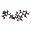  Type: oligosaccharide, Oligosaccharide / Class: Inhibitor / Mass: 645.606 Da / Num. of mol.: 1 Type: oligosaccharide, Oligosaccharide / Class: Inhibitor / Mass: 645.606 Da / Num. of mol.: 1Source method: isolated from a genetically manipulated source Details: oligosaccharide / References: alpha-acarbose |
|---|---|
| #3: Sugar | ChemComp-GLC /  Type: D-saccharide, alpha linking / Mass: 180.156 Da / Num. of mol.: 1 Type: D-saccharide, alpha linking / Mass: 180.156 Da / Num. of mol.: 1Source method: isolated from a genetically manipulated source Formula: C6H12O6 |
| #4: Sugar |  Type: D-saccharide / Mass: 321.324 Da / Num. of mol.: 2 Type: D-saccharide / Mass: 321.324 Da / Num. of mol.: 2Source method: isolated from a genetically manipulated source Formula: C13H23NO8 |
-Non-polymers , 2 types, 517 molecules 

| #5: Chemical | ChemComp-GOL /  Mass: 92.094 Da / Num. of mol.: 1 / Source method: obtained synthetically / Formula: C3H8O3 Mass: 92.094 Da / Num. of mol.: 1 / Source method: obtained synthetically / Formula: C3H8O3 |
|---|---|
| #6: Water | ChemComp-HOH / Mass: 18.015 Da / Num. of mol.: 516 / Source method: isolated from a natural source / Formula: H2O |
-Experimental details
-Experiment
| Experiment | Method: X-RAY DIFFRACTION / Number of used crystals: 1 |
|---|
- Sample preparation
| Crystal | Density Matthews: 2.31 Å3/Da / Density % sol: 46.82 % |
|---|---|
| Crystal grow | Temperature: 289 K / Method: vapor diffusion, sitting drop / pH: 5.5 Details: 0.1M BIS-TRIS, 25% PEG3350, 10% ACARBOSE, pH 5.5, VAPOR DIFFUSION, SITTING DROP, temperature 289K |
-Data collection
| Diffraction | Mean temperature: 100 K |
|---|---|
| Diffraction source | Source: SYNCHROTRON / Site: APS  / Beamline: 19-ID / Wavelength: 0.97921 Å / Beamline: 19-ID / Wavelength: 0.97921 Å |
| Detector | Type: ADSC QUANTUM 315r / Detector: CCD / Date: Apr 22, 2010 / Details: MIRROR |
| Radiation | Monochromator: SI 111 CRYSTAL / Protocol: SINGLE WAVELENGTH / Monochromatic (M) / Laue (L): M / Scattering type: x-ray |
| Radiation wavelength | Wavelength: 0.97921 Å / Relative weight: 1 |
| Reflection | Resolution: 1.99→42 Å / Num. all: 91803 / Num. obs: 91803 / % possible obs: 96 % / Observed criterion σ(F): 0 / Observed criterion σ(I): 0 / Redundancy: 3.4 % / Rmerge(I) obs: 0.103 / Net I/σ(I): 12.2 |
| Reflection shell | Resolution: 2→2.03 Å / Redundancy: 2.7 % / Rmerge(I) obs: 0.585 / Mean I/σ(I) obs: 1.16 / % possible all: 84.9 |
- Processing
| Software |
| |||||||||||||||||||||||||||||||||||||||||||||||||||||||||||||||||||||||||||||
|---|---|---|---|---|---|---|---|---|---|---|---|---|---|---|---|---|---|---|---|---|---|---|---|---|---|---|---|---|---|---|---|---|---|---|---|---|---|---|---|---|---|---|---|---|---|---|---|---|---|---|---|---|---|---|---|---|---|---|---|---|---|---|---|---|---|---|---|---|---|---|---|---|---|---|---|---|---|---|
| Refinement | Method to determine structure: MOLECULAR REPLACEMENT Starting model: PDB ENTRY 3N04 Resolution: 1.99→41.365 Å / SU ML: 0.27 / σ(F): 0 / Phase error: 28.24 / Stereochemistry target values: ML
| |||||||||||||||||||||||||||||||||||||||||||||||||||||||||||||||||||||||||||||
| Solvent computation | Shrinkage radii: 0.95 Å / VDW probe radii: 1.2 Å / Solvent model: FLAT BULK SOLVENT MODEL / Bsol: 39.417 Å2 / ksol: 0.321 e/Å3 | |||||||||||||||||||||||||||||||||||||||||||||||||||||||||||||||||||||||||||||
| Displacement parameters |
| |||||||||||||||||||||||||||||||||||||||||||||||||||||||||||||||||||||||||||||
| Refinement step | Cycle: LAST / Resolution: 1.99→41.365 Å
| |||||||||||||||||||||||||||||||||||||||||||||||||||||||||||||||||||||||||||||
| Refine LS restraints |
| |||||||||||||||||||||||||||||||||||||||||||||||||||||||||||||||||||||||||||||
| LS refinement shell |
| |||||||||||||||||||||||||||||||||||||||||||||||||||||||||||||||||||||||||||||
| Refinement TLS params. | Method: refined / Refine-ID: X-RAY DIFFRACTION
| |||||||||||||||||||||||||||||||||||||||||||||||||||||||||||||||||||||||||||||
| Refinement TLS group |
|
